The human spine is a biomechanical masterpiece of competing evolutionary demands: it must provide rigid, load-bearing structural support to balance the torso in upright bipedal posture, while simultaneously preserving multi-planar flexibility and encasing the delicate neural elements of the spinal cord and cauda equina.
Every day, the lumbar spine bears extraordinary compressive, shear, and torsional forces. During a simple standing forward bend while lifting a modest 20-kilogram weight, the compressive force concentrated across the L4�L5 intervertebral disc exceeds 3,000 Newtons�equivalent to the weight of an adult male grizzly bear pressing directly upon a fibrocartilaginous disc measuring less than five centimeters in diameter.
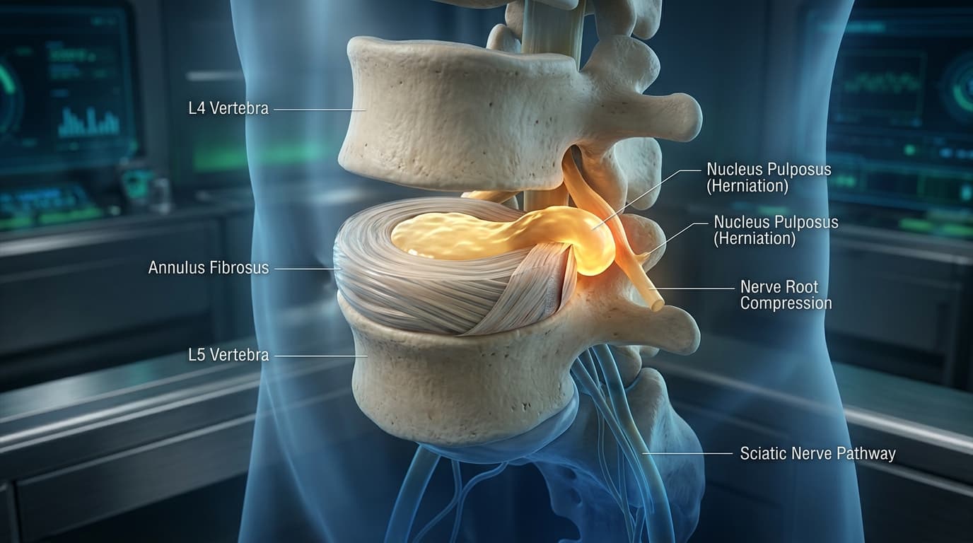
When repetitive mechanical overload or age-related biochemical desiccation degrades the structural integrity of the disc's outer fibrous envelope, the pressurized internal gel herniates posteriorly, triggering severe, disabling radiculopathy: Sciatica.
In this clinical masterclass, we explore the articulated 3D anatomy of the vertebral column, deconstruct the multi-lamellar microstructure of the intervertebral disc, analyze Nachemson intradiscal pressure variations, and trace the mechanical and neurochemical pathways of sciatic nerve root compression using our interactive 3D WebGL workstation.
Interactive 3D Studio: Simulate L4-L5 Disc Herniation
Run exact formula simulations on NexProTools.
1. The Articulated Vertebral Column: Curvatures & Kinematic Units
The adult human spinal column comprises 26 distinct articulated osseous segments organized into four physiological sagittal curvatures:
| Spinal Curvature Region | Vertebral Segments | Curve Orientation | Biomechanical Function | | :--- | :--- | :--- | :--- | | **Cervical Lordosis** | C1 - C7 | Convex Anteriorly | Balances cranium (approx. 5 kg) over thorax with minimal muscular torque | | **Thoracic Kyphosis** | T1 - T12 | Concave Anteriorly | Accommodates intrathoracic cardiopulmonary volume; rib cage stabilization | | **Lumbar Lordosis** | L1 - L5 | Convex Anteriorly | Transmits trunk load to pelvis; cushions bipedal ground reaction shock waves | | **Sacrococcygeal Kyphosis**| S1 - Co4 | Concave Anteriorly | Pelvic ring attachment; distributes weight across sacroiliac joints |
The Biomechanical Value of Sagittal Curvature
The alternating lordotic and kyphotic curves are not anatomical imperfections; they are critical shock-absorbing adaptations. According to the classic Euler-Delpech Resistance Formula, the structural resistance of a curved column is proportional to the square of its curves plus one:
Where N is the number of physiological spinal curves:
- A straight column (
N = 0) has a resistance factor ofR = 1. - The human spine with its three mobile physiological curves (
N = 3: Cervical, Thoracic, Lumbar) has a resistance factor ofR = 3� + 1 = 10. - The curved human spine is ten times more resistant to axial compressive shock loads than a straight, uncurved osseous rod.
The Functional Spinal Unit (FSU) of Junghanns
The smallest physiological kinematic motion segment of the spine is the Functional Spinal Unit (FSU), consisting of:
- Two adjacent vertebral bodies.
- The intervening intervertebral disc.
- The bilateral posterior synovial Zygapophysial (Facet) Joints.
- The stabilizing interconnecting ligaments: Anterior Longitudinal Ligament (
ALL), Posterior Longitudinal Ligament (PLL), Ligamentum Flavum, Interspinous, and Supraspinous ligaments.
| Vertebral Component | Anatomical Feature | Biomechanical Role in Functional Spinal Unit | | :--- | :--- | :--- | | **Vertebral Body** | Massive kidney-shaped trabecular core | Bears 80% - 85% of axial compressive trunk loads | | **Pedicles** | Short, thick rounded osseous bridges | Channels anterior axial force to posterior neural arch | | **Transverse Processes** | Slender lateral levers | Moment arms for psoas, quadratus lumborum, and deep paraspinal rotators | | **Laminae & Facet Joints** | Synovial zygapophysial joints (Sagittal orientation) | Permits sagittal flexion-extension, strictly resists rotational axial torsion | | **Spinous Process** | Thick, quadrangular hatchet-shaped blade | Anchors erector spinae and interspinous ligaments | | **Vertebral Foramen** | Trefoil triangular neural canal | Encapsulates dural thecal sac and cauda equina roots |
The Three-Column Spine Model of Denis
In spinal trauma and biomechanics, stability is assessed through the Denis Three-Column Model:
- Anterior Column: Anterior half of the vertebral body, anterior half of the intervertebral disc, and the dense Anterior Longitudinal Ligament (
ALL). - Middle Column: Posterior half of the vertebral body, posterior half of the disc, and the Posterior Longitudinal Ligament (
PLL). Disruption of the middle column is the defining criterion of clinical mechanical spinal instability. - Posterior Column: Pedicles, laminae, facet joints, spinous processes, ligamentum flavum, and interspinous ligaments.
2. The Intervertebral Disc: Annulus Fibrosus vs Nucleus Pulposus
The intervertebral disc is the largest avascular organ in the human body. It functions as a hydraulic shock absorber, transforming axial compressive loads into radial tensile hoop stress.
``> CONCENTRIC MICROSTRUCTURAL LAYERS OF THE LUMBAR DISC
- Outer Annulus Fibrosus: 15 to 25 concentric lamellae of dense Type I Collagen running at alternating $+30�$ and $-30�$ angles to contain circumferential hoop stress.
- Inner Annulus Fibrosus: Fibrocartilaginous transitional zone enriched with Type II collagen and chondrocytes.
- Central Nucleus Pulposus: Hydrophilic proteoglycan gel (80% water bound to aggrecan) distributing hydraulic pressure isotropically.
- Cartilaginous Endplates: Superior and inferior hyaline cartilage layers through which essential nutrients diffuse into the avascular disc.``
| Biochemical Component | Annulus Fibrosus | Nucleus Pulposus | | :--- | :--- | :--- | | **Primary Cell Type** | Fibroblast-like elongated cells | Chondrocyte-like spherical cells | | **Water Content (Youth)** | 65% - 70% | 80% - 88% (Highly hydrated gel) | | **Water Content (Elderly)**| 55% - 60% | 65% - 70% (Biochemically desiccated) | | **Dominant Collagen Type** | **Type I Collagen (80%)** | **Type II Collagen (80%)** | | **Proteoglycan Concentration**| Low (Aggrecan ~ 15% dry weight) | **Extreme (Aggrecan ~ 65% dry weight)** | | **Primary Biomechanical Role**| Resists tensile circumferential hoop stress | Converts compressive loads into isotropic fluid pressure |
The Nucleus Pulposus: The Pressurized Gel Core
Positioned slightly posterior to the center in the lumbar spine, the nucleus pulposus is a gelatinous, hydrophilic matrix:
- Aggrecan & Glycosaminoglycans (
GAGs): Packed with negatively charged chondroitin sulfate and keratan sulfate side chains. - Fixed Charge Density (
FCD): These dense negative charges create a high interstitial osmotic pressure (Donnan equilibrium), drawing water into the disc. In healthy young adults, this osmotic swelling pressure generates an intrinsic resting baseline pressure of0.1to0.2 MPa, keeping the disc taut even when lying down. - Under axial loading, the incompressible water cannot escape rapidly through the dense matrix. Instead, the nucleus acts as an isotropic fluid, transmitting pressure equally in all radial directions outward against the surrounding ring.
The Annulus Fibrosus: The Tensile Containment Belt
Surrounding the nucleus is the annulus fibrosus, composed of 15 to 25 concentric fibrous sheets (Lamellae):
- Alternating Ply Architecture: The dense Type I collagen fibers within each lamella run at an angle of roughly
30�relative to the vertebral endplate. Crucially, the fibers in adjacent lamellae run in alternating directions (+30�and-30�). - Mechanical Significance: This cross-ply criss-cross arrangement is mechanically identical to modern radial automobile tires. It resists torsional twisting forces (half the fibers tighten during rotation in either direction) and converts the hydrostatic pressure of the expanding nucleus into circumferential Hoop Stress.
3. Biomechanics of Intradiscal Pressure: The Nachemson Evidence
In landmark in vivo orthopedic studies pioneered by Alf Nachemson and confirmed by Wilke et al., miniature pressure transducers were inserted directly into the L3�L4 and L4�L5 nucleus pulposus of human volunteers during various activities.
The findings completely transformed modern spinal ergonomics:
| Posture / Physical Activity | Relative Intradiscal Pressure (% of Standing) | Biomechanical Loading Mechanism | | :--- | :--- | :--- | | **Lying Supine (Flat on Back)** | **25%** | Minimal axial gravity; osmotic rehydration occurs | | **Lying on Side (Lateral Decubitus)**| **75%** | Mild muscle tone maintains baseline tension | | **Standing Upright (Normal Lordosis)** | **100% (Baseline Reference)** | Even load sharing between disc (80%) and facet joints (20%) | | **Walking at Moderate Pace** | **115%** | Dynamic cyclical ground reaction impulse | | **Sitting Upright (Ergonomic 110�)** | **140%** | Mild posterior pelvic tilt increases disc shear | | **Sitting Slouched (Forward Kyphosis)**| **185%** | Pelvis tilts backward; wedges disc posteriorly against PLL | | **Standing & Bending Forward (30� Flexion)**| **150%** | Trunk center of mass moves anterior to spine axis | | **Bending Forward while Lifting 20 kg**| **275% - 350%** | Extreme lever arm creates >3,000 N compressive barotrauma |
Why Slouched Sitting Generates Higher Disc Pressure than Standing
Many individuals assume sitting is restful for the back. Biomechanically, the opposite is true:
- When standing, the pelvis tilts naturally anteriorly, preserving lumbar lordosis. The posterior facet joints share roughly 15% to 25% of the axial compressive load, shielding the intervertebral disc.
- When sitting without lumbar support, the pelvis tilts posteriorly. This forces the lumbar spine to flatten its natural lordosis and enter flexion (kyphosis).
- In lumbar flexion, the anterior margins of the vertebral bodies tilt toward each other, compressing the anterior annulus like a wedge.
- This wedging forces the pressurized nucleus pulposus posteriorly against the thinner, weaker posterior annulus fibrosus, driving intradiscal pressure from
100%up to185%.
4. The Pathophysiology of Lumbar Disc Herniation
Lumbar disc herniation is rarely an instantaneous single-event injury in an otherwise healthy disc; it is the culmination of progressive biochemical desiccation, micro-fissuring, and mechanical fatigue.
``> THE FOUR CLINICAL STAGES OF DISC HERNIATION (NASS CONSENSUS)
- Stage 1: Internal Degeneration: Desiccation of nuclear aggrecan leads to microscopic radial fissures within inner annular lamellae.
- Stage 2: Protrusion (Prolapse / Bulge): Nuclear material migrates along fissures; outer annulus and posterior longitudinal ligament remain intact, forming a broad-based bulge.
- Stage 3: Extrusion: Complete tear through all annular lamellae; pressurized nuclear gel spills into the epidural space but remains contiguous with parent disc.
- Stage 4: Sequestration (Free Fragment): Extruded nuclear fragment detaches completely, migrating superiorly or inferiorly to lodge against the traversing nerve root.``
The 4 Morphological Stages of Herniation
According to international North American Spine Society (NASS) consensus classifications:
Stage 1: Disc Degeneration & Annular Fissuring
Age-related loss of chondroitin sulfate side chains reduces the fixed charge density of the nucleus pulposus. The disc desiccates (loses water), dropping from 85% water content in youth to below 65%. The desorbed nucleus can no longer distribute hydraulic loads isotropically. Focal mechanical stress concentrations cause microscopic circumferential tears within the inner annular lamellae, coalescing into full-thickness Radial Fissures.
Stage 2: Disc Protrusion (Prolapse / Bulge)
The dehydrated nucleus pulposus migrates along a radial fissure toward the outer margin of the disc. The outer lamellae of the annulus fibrosus and the overlying Posterior Longitudinal Ligament (PLL) remain physically intact, but bulge outward into the spinal canal. The base of the herniated material is wider than any apex extension.
Stage 3: Disc Extrusion
The pressurized nucleus pulposus ruptures completely through all outer lamellae of the annulus fibrosus. The extruded gelatinous mass extends into the epidural space through a narrow defect, but remains contiguous with the parent disc tissue within the interspace.
Stage 4: Disc Sequestration (Free Fragment)
The extruded nucleus pulposus fragment completely breaks off from the parent disc. The free sequestered fragment migrates superiorly or inferiorly within the epidural space along the anterior or posterior aspect of the thecal sac, frequently lodging within the subarticular lateral recess where it directly impinges upon the traversing spinal nerve root.
| Anatomical Quadrant | Ligamentous Reinforcement | Annular Lamellar Thickness | Herniation Vulnerability | | :--- | :--- | :--- | :--- | | **Anterior Quadrant** | Thick, dense Anterior Longitudinal Ligament (ALL) | Broad, densely packed Type I collagen | **Extremely Rare (< 1%)** | | **Posterior Midline** | Midline band of Posterior Longitudinal Ligament (PLL) | Moderate lamellar density | **Infrequent (~5%)** (Can cause cauda equina syndrome if massive) | | **Posterior-Lateral Recess**| **PLL thins by 50% at L4-L5, leaving corner uncovered** | **Thinnest annular lamellar boundary** | **Predominant Site (> 90%) (Compreses traversing sciatic nerve root)** | | **Far Lateral (Foraminal)**| Intertransverse ligaments | Thinned outer border | **Uncommon (~5%)** (Compresses exiting nerve root) |
Why Herniations Occur Posterior-Laterally
Over 90% of all symptomatic lumbar disc herniations occur at L4�L5 or L5�S1, and specifically in the posterior-lateral quadrant:
- PLL Thinning: The Anterior Longitudinal Ligament (
ALL) is broad, thick, and extraordinarily strong, completely shielding the anterior annulus. In contrast, the Posterior Longitudinal Ligament (PLL) narrows progressively as it descends down the lumbar spine, measuring only half its thoracic width at L4 and L5, leaving the posterior-lateral aspect of the disc completely uncovered. - Posterior Annular Thinning: The posterior lamellae of the annulus fibrosus are biologically thinner and contain fewer tightly bound cross-links than the anterior lamellae.
- Flexion Mechanics: Daily activities (bending forward, lifting, slouched sitting) consistently place the lumbar spine in flexion, forcing the nuclear gel posteriorly.
5. Nerve Root Anatomy & The Sciatica Mechanism: Mechanical vs Chemical
Sciatica (lumbar radiculopathy) is severe radiating pain originating in the lower back or buttock and shooting down the posterior or lateral thigh, calf, and foot along the anatomical distribution of the sciatic nerve.
Historically, clinicians believed sciatica was caused purely by direct mechanical compression of the nerve root by the bulging disc. Modern neurobiology has revealed that sciatica is a dual mechanical and biochemical inflammatory disease.
| Pathological Hit | Primary Etiology | Biological Cascade | Clinical Symptom Manifestation | | :--- | :--- | :--- | :--- | | **Hit 1: Mechanical Ischemia** | Direct physical compression by extruded nuclear fragment against pedicle | Venous stasis ( - 20\text{ mmHg}$), capillary arrest ( - 50\text{ mmHg}$), axonal ischemia | Conduction block: numbness, paresthesias, motor weakness (Foot Drop) | | **Hit 2: Chemical Inflammation**| Exposure of immunologically privileged nucleus pulposus to epidural space | High Phospholipase A2 ($) and -\alpha$ provoke intense epineurial chemical burn | Severe intractable shooting radicular leg pain, allodynia, hyperalgesia |
Hit 1: The Chemical Inflammatory Cascade
The interior of the nucleus pulposus is an immunologically privileged site. Because the adult disc is entirely avascular, the body's immune system has never encountered nucleus pulposus antigens during fetal development:
- When a disc extrudes into the vascularized epidural space, the immune system recognizes the nuclear gel as foreign tissue.
- The nucleus contains extraordinary concentrations of Phospholipase A2 (
PLA2)�over 10,000 times higher than that found in any other human tissue. PLA2hydrolyzes cell membrane phospholipids, initiating the arachidonic acid cascade to flood the local space with pro-inflammatory Prostaglandin E2 (PGE2), Interleukin-1\beta(IL-1\beta), and Tumor Necrosis Factor-\alpha(TNF-\alpha).- These cytokines cause an intense chemical burn of the unmyelinated epineurium, dramatically lowering the firing threshold of nociceptive C-fibers and inducing extreme hyperalgesia even in the absence of severe physical compression.
Hit 2: The Mechanical Microvascular Strangulation
Spinal nerve roots differ fundamentally from peripheral nerves: they lack a thick, protective perineurium and rely on a delicate capillary plexus. When an extruded disc fragment compresses a nerve root against the bony pedicle or facet:
- Low pressure (
10 - 20 mmHg): Compresses the thin-walled intraradicular veins, causing venous stasis, local capillary thrombosis, and endoneurial edema. - Moderate pressure (
30 - 50 mmHg): Compromises capillary arterial perfusion, producing axonal ischemia and axonal transport arrest. - High pressure (
> 75 mmHg): Blocks electrical action potential propagation, manifesting clinically as numbness, paresthesias, and progressive motor weakness (foot drop).
6. Clinical Neurological Diagnostics: Dermatomes, Myotomes & Reflexes
Determining the exact anatomical level of a lumbar disc herniation during a bedside physical examination relies on precise evaluation of the three lower lumbar and sacral nerve roots: L4, L5, and S1.
| Spinal Nerve Root | Primary Myotome (Motor Testing) | Sensory Dermatome Distribution | Reflex Arc Assessment | | :--- | :--- | :--- | :--- | | **L4 Nerve Root** | Quadriceps (Knee extension), Tibialis Anterior (Ankle inversion) | Medial lower calf, anterior knee, and medial malleolus | **Patellar Tendon Reflex (Diminished / Absent)** | | **L5 Nerve Root** | Extensor Hallucis Longus (Great toe dorsiflexion), Gluteus Medius | Anterolateral leg, dorsum of foot, and web space of 1st-2nd toes | **None (No reliable deep tendon reflex arc)** | | **S1 Nerve Root** | Gastrocnemius / Soleus (Ankle plantarflexion), Peroneus Longus | Lateral border of foot, plantar heel, and small 5th toe | **Achilles Tendon Reflex (Diminished / Absent)** |
The Traversing vs Exiting Nerve Root Rule
In the lumbar spine, nerve roots exit through the neural foramina beneath their corresponding pedicles. Because lumbar roots travel vertically downward before exiting:
- An L4�L5 Paracentral Disc Herniation (the classic 90% presentation) does NOT compress the exiting L4 nerve root; it compresses the Traversing L5 Nerve Root as it crosses the disc space to exit below at L5�S1.
- An L5�S1 Paracentral Disc Herniation compresses the Traversing S1 Nerve Root.
- Exception: A far-lateral (foraminal) disc herniation directly compresses the Exiting Nerve Root at that level (e.g. far-lateral L4�L5 herniation compresses L4).
Bedside Provocative Tests
- Straight Leg Raise (
SLR/ Las�gue Sign): With the patient supine, the clinician passively elevates the fully extended leg. A positive test elicits sharp, electric shooting pain radiating below the knee between30�and70�of elevation, confirming mechanical tension on the L5 or S1 nerve roots as they glide across the herniated fragment. - Crossed Straight Leg Raise (Fajersztajn Test): Raising the unaffected, asymptomatic leg reproduces severe radiating pain down the affected, symptomatic leg. Highly specific (
> 90%) for a large extruded or sequestered free fragment.
7. Interactive 3D Disassembly & Herniation Simulation Protocol
To visualize these kinetic principles on our 3D Human Anatomy Studio, follow this structured four-step clinical exploration protocol:
Step 1: Vertebral Column Separation (Exploded View: 0% to 50%)
- Rotate the articulated spine model to view the anterior lumbar vertebral bodies (L3, L4, L5) and the sacral base.
- Drag the Layer Disassembly Slider to
35%. - Observe the vertical separation of the vertebrae along the axial column:
- Notice how the intervertebral disc endplates unseat from the vertebral bodies.
- Inspect the posterior facet joints (superior and inferior articular processes) disengaging.
- Trace the central yellow spinal cord (thecal sac) running through the neural canal.
Step 2: Posterior Element & Canal Inspection
- Rotate the camera to look down the spinal canal from a superior-posterior perspective.
- Locate the exiting bilateral yellow spinal nerve roots emerging at each vertebral level beneath the pedicles.
- Observe how close the nerve roots course relative to the posterior-lateral margin of the intervertebral discs.
Step 3: Disc Microstructure Examination
- Focus the view directly on the L4�L5 Intervertebral Disc.
- Identify the outer grey concentric rings representing the multi-lamellar Annulus Fibrosus.
- Identify the inner cyan-tinted core representing the hydrated Nucleus Pulposus.
Step 4: Pathology Mode Activation (L4�L5 Herniation Simulation)
- Toggle Clinical Pathology Simulation to
Active. - Notice the procedural formation of a posterior-lateral tear in the L4�L5 annulus.
- Drag the Severity Slider from
20%to90%:- Watch the extruded red/orange nucleus pulposus mass emerge through the annular fissure.
- Observe the expanding herniation fragment press directly against the descending right L5 spinal nerve root.
- Watch the nerve root shift from normal amber to bright inflamed red, visually demonstrating the mechanical impingement and neurochemical inflammation that produces clinical Sciatica.
8. Summary & Key Takeaways
CLINICAL TAKEAWAYS: 3D SPINE BIOMECHANICS & HERNIATION
- The 3 Sagittal Curves of the human spine increase structural axial resistance by a factor of 10 (
R = N� + 1 = 10) compared to a straight column.- The Intervertebral Disc functions as a cross-ply composite: the pressurized, hydrophilic nucleus pulposus distributes hydraulic loads isotropically, while alternating
30�Type I collagen lamellae in the annulus fibrosus contain tensile hoop stress.- Slouched Sitting dramatically elevates L4�L5 intradiscal pressure to 185% of standing baseline, wedging the nucleus posteriorly against the thinned Posterior Longitudinal Ligament.
- Sciatica is a Dual Hit: direct mechanical microvascular ischemia coupled with intense chemical inflammatory radiculitis driven by exposed nuclear Phospholipase A2 (
PLA2) andTNF-\alpha.- An L4�L5 Paracentral Herniation typically compresses the traversing L5 Nerve Root, manifesting as great toe extensor weakness (foot drop), numbness over the foot dorsum, and a positive Straight Leg Raise.
Inspect these biomechanical forces in real-time WebGL on our Interactive 3D Human Anatomy Workstation.