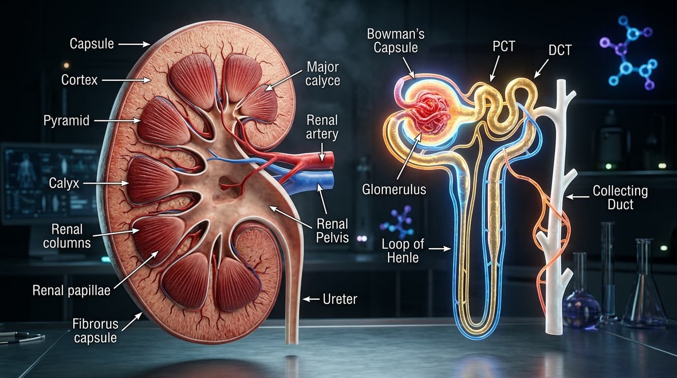
The human kidneys comprise less than 0.5% of total body weight, yet they consume over 20% to 25% of resting cardiac output�receiving approximately 1,200 milliliters of whole blood per minute. Within each kidney, an intricate network of 1.0 to 1.2 million microscopic functional filtration units, termed Nephrons, continuously filters roughly 180 liters of blood plasma per day. From this immense volume of ultrafiltrate, the nephron reabsorbs more than 99% of filtered water and sodium, concentrating metabolic wastes into 1.5 liters of excreted urine.
Understanding renal medicine requires linking macroscopic organ morphology�such as the corticomedullary osmolar gradient�with the sub-microscopic physics of glomerular capillary Starling forces and podocyte slit diaphragm permeability.
In this clinical masterclass, we deconstruct the gross macroscopic architecture of the human kidney, zoom into the high-resolution 3D anatomy of the nephron, derive the mathematical equations governing glomerular filtration, and analyze the cellular pathophysiology of diabetic glomerulosclerosis using our interactive 3D WebGL workstation.
Interactive 3D Studio: Inspect 3D Nephron Filtration
Run exact formula simulations on NexProTools.
1. Macroscopic Kidney Architecture: Cortex, Medulla & Vascular Hilum
The kidneys are bilateral, retroperitoneal bean-shaped organs positioned against the posterior abdominal wall on either side of the vertebral column, extending from the level of T12 to L3. Because the liver displaces abdominal viscera on the right, the right kidney sits approximately 1.5 cm to 2.0 cm lower than the left.
| Anatomical Metric | Adult Normal Range | Clinical Significance | | :--- | :--- | :--- | | **Bipolar Renal Length** | 10.0 - 12.5 cm | < 9.0 cm indicates chronic irreversible nephron atrophy (CKD) | | **Renal Width (Transverse)** | 5.0 - 6.5 cm | Essential for total renal parenchymal volume estimation | | **Renal Thickness (AP)** | 3.0 - 3.8 cm | Anteroposterior diameter on cross-sectional CT/MRI | | **Cortical Thickness** | 8.0 - 10.5 mm | Cortical thinning (< 7mm) is a hallmark of global sclerosis | | **Adult Kidney Weight** | 135 - 170 grams | Strongly correlates with baseline functional nephron endowment | | **Renal Blood Flow (RBF)** | 1,000 - 1,250 mL/min | Consumes 20% - 25% of total resting cardiac output |
The Three Concentric Parenchymal Zones
A longitudinal coronal cross-section through the human kidney reveals three sharply demarcated anatomical zones:
``> ANATOMICAL CONCENTRIC ZONES OF THE HUMAN KIDNEY
- Fibrous Renal Capsule: Tough collagenous protective envelope resisting interstitial swelling.
- Renal Cortex: Granular outer mantle housing 100% of all 1.2 million glomeruli and convoluted tubules (PCT & DCT).
- Renal Medulla: 8 to 18 striated conical pyramids housing parallel loops of Henle, collecting ducts, and the vasa recta.
- Collecting Funnel: Renal Papillae $\to$ Minor Calyces $\to$ Major Calyces $\to$ Infundibular Renal Pelvis $\to$ Ureter.``
1. The Renal Fibrous Capsule
A tough, collagenous protective envelope that resists intraparenchymal swelling. Because the non-compliant capsule does not stretch easily, acute acute inflammation (e.g. acute pyelonephritis) or rapid urinary tract obstruction leads to extreme interstitial pressure elevations, triggering severe visceral renal colic.
2. The Renal Cortex
The pale, granular outer zone extending from the fibrous capsule to the base of the medullary pyramids, as well as projecting between the pyramids as the Renal Columns of Bertin:
- Contains 100% of all renal corpuscles (glomerular capillary tufts and Bowman's capsules).
- Houses the high-metabolism convoluted segments: Proximal Convoluted Tubules (
PCT) and Distal Convoluted Tubules (DCT). - Normal cortical thickness measures
8.0 mmto10.5 mm. In progressive Chronic Kidney Disease (CKD), cortical parenchymal loss produces marked cortical thinning on ultrasound (< 7.0 mm), reflecting irreversible global nephron loss.
3. The Renal Medulla
Composed of 8 to 18 striated, conical Renal Pyramids whose bases face the outer cortex and whose apices (the Renal Papillae) project inward toward the minor calyces:
- Consists exclusively of straight parallel tubular structures: the descending and ascending limbs of the Loop of Henle, collecting ducts, and the hairpin vascular loops of the Vasa Recta.
- Maintains an extreme vertical osmotic gradient (from
300 mOsm/kgat the corticomedullary junction to over1,200 mOsm/kgin the deep inner papilla), essential for concentrating urine under the control of antidiuretic hormone (ADH / Vasopressin).
2. Microscopic 3D Nephron Architecture: The Tubular Continuum
The nephron is the functional epithelial tube that transforms glomerular ultrafiltrate into finished urine. Structurally, nephrons are classified into two distinct populations based on the location of their glomeruli:
| Structural Feature | Cortical Nephrons (85% of total) | Juxtamedullary Nephrons (15% of total) | | :--- | :--- | :--- | | **Glomerular Location** | Outer and mid-cortex | Deep cortex immediately adjacent to corticomedullary junction | | **Loop of Henle Length** | Short (Reaches only outer medulla) | Long hairpin loops (Penetrates deep inner medullary papillae) | | **Peritubular Blood Supply**| Standard capillary network | Specialized hairpin loops (**Vasa Recta**) | | **Primary Functional Role** | Bulk solute and water reabsorption (PCT) | Generation of hypertonic osmolar gradient for urine concentration | | **Juxtaglomerular Renin** | Lower renin granule density | Dense renin secretion regulating systemic hemodynamics |
| Nephron Pipeline Stage | Predominant Epithelium | Key Transport Machinery | Fraction Filtered Load Reabsorbed | | :--- | :--- | :--- | :--- | | **1. Bowman Capsule & Glomerulus** | Podocytes & fenestrated endothelium | Size/charge selective barrier | Produces 180 L/day of ultrafiltrate | | **2. Proximal Convoluted Tubule** | Cuboidal with dense brush border | SGLT2, Na+/H+ exchanger (NHE3) | 65% - 70% Na+ & H2O, 100% glucose & amino acids | | **3. Descending Thin Limb of Henle** | Simple squamous epithelium | High-density Aquaporin-1 (AQP1) | 15% H2O (Permeable to water, impermeable to NaCl) | | **4. Thick Ascending Limb (TAL)** | Cuboidal, no brush border, dense mito| Apical NKCC2 co-transporter | 20% - 25% Na+/K+/Cl- (Impermeable to water) | | **5. Distal Convoluted Tubule** | Tight cuboidal epithelium | Thiazide-sensitive NCC co-transporter| 5% Na+/Cl- fine-tuning | | **6. Cortical & Medullary Collecting**| Principal & intercalated cells | ENaC, ROMK, ADH-regulated AQP2 | Final 1% - 4% water and electrolyte excretion control |
Stage 1: The Glomerulus & Bowman's Capsule
The vascular pole of the nephron where arterial filtration begins. Blood enters via the Afferent Arteriole into a high-pressure, non-anastomosing network of 20 to 40 specialized capillary loops (the glomerular tuft), then exits via the Efferent Arteriole. Surrounding this capillary tuft is Bowman's Capsule, a double-walled cup composed of an inner visceral layer (specialized podocytes) and an outer parietal epithelial layer enclosing Bowman's Space.
Stage 2: Proximal Convoluted Tubule (PCT)
Originating from the urinary pole of Bowman's capsule, the PCT is the energetic workhorse of the nephron:
- Lined by simple cuboidal epithelial cells with dense apical microvilli forming a prominent Brush Border, which increases luminal surface area by more than 20-fold.
- Packed with densely arrayed basolateral mitochondria driving
Na+/K+-ATPasepumps. - Responsible for the obligate reabsorption of 65% to 70% of filtered water and sodium, 100% of filtered glucose and amino acids (via
SGLT2and sodium-amino acid symporters), and 85% of filtered bicarbonate.
Stage 3: The Loop of Henle & Counter-Current Multiplication
- Thin Descending Limb: Extremely permeable to water via high-density aquaporin-1 (
AQP1) channels, but virtually impermeable to sodium and urea. As filtrate descends into the hypertonic medullary interstitium, water leaves the lumen, concentrating luminal fluid up to1,200 mOsm/kgat the hairpin turn. - Thick Ascending Limb (
TAL): Structurally and functionally the inverse of the descending limb. The TAL is completely impermeable to water, but actively pumps electrolytes out of the tubular lumen into the medullary interstitium via the apicalNa+/K+/2Cl-co-transporter (NKCC2). This uncoupling of water reabsorption from solute transport dilutes the luminal fluid down to100 mOsm/kg(hypotonic) while depositing salt into the medullary interstitium.
Clinical Pharmacological Insight: The NKCC2 co-transporter in the thick ascending limb is the specific molecular target of loop diuretics (Furosemide, Bumetanide, Torsemide). By blocking NKCC2, these agents abolish the medullary hypertonic gradient, preventing downstream water reabsorption and inducing potent naturesis.
3. Glomerular Hemodynamics & Starling Ultrafiltration Forces
Glomerular ultrafiltration is governed by the classic Starling equation of transcapillary fluid exchange. Unlike systemic skeletal muscle capillaries�where filtration occurs at the arteriolar end and reabsorption occurs at the venular end�glomerular capillaries maintain net positive filtration along their entire length.
Where:
K_fis the ultrafiltration coefficient (the product of capillary surface area and hydraulic water permeability, normally\approx 12.5\ \text{mL/min/mmHg}).P_{GC}is Glomerular Capillary Hydrostatic Pressure (\approx 50\ \text{mmHg}), favoring filtration.P_{BS}is Bowman's Space Hydrostatic Pressure (\approx 12\ \text{mmHg}), opposing filtration.\pi_{GC}is Glomerular Capillary Oncotic Pressure (\approx 28 - 35\ \text{mmHg}), opposing filtration.\pi_{BS}is Bowman's Space Oncotic Pressure (\approx 0\ \text{mmHg}under healthy conditions, as proteins are excluded).
| Starling Pressure Parameter | Symbol | Afferent Arteriolar End | Efferent Arteriolar End | Physiological Determinant | | :--- | :--- | :--- | :--- | :--- | | **Capillary Hydrostatic Pressure** | P_GC | **50 mmHg** | **48 mmHg** | Favors filtration; sustained by downstream efferent resistance | | **Bowman Space Hydrostatic Pressure** | P_BS | **12 mmHg** | **12 mmHg** | Opposes filtration; elevates sharply during urinary obstruction | | **Capillary Plasma Oncotic Pressure** | pi_GC | **28 mmHg** | **35 mmHg** | Opposes filtration; rises as water is filtered and protein concentrates | | **Bowman Space Oncotic Pressure** | pi_BS | **0 mmHg** | **0 mmHg** | Effectively zero because healthy GBM excludes plasma proteins | | **NET ULTRAFILTRATION PRESSURE** | **Net Delta P** | **+10 mmHg (Filtration)** | **+1 mmHg (Filtration)** | **Net positive filtration pressure along entire capillary length** |
The Dual-Arteriole Pressure Valve: Afferent vs Efferent Tone
A unique anatomical feature of the renal circulation is that the glomerular capillary bed is positioned between two resistance vessels: the Afferent and Efferent arterioles. This allows precise regulation of both Renal Blood Flow (RBF) and Glomerular Filtration Rate (GFR):
``> VASCULAR RESISTANCE MODULATION OF GLOMERULAR FILTRATION
- Afferent Arteriolar Dilation (Mediated by Prostaglandins PGE2 / PGI2, Nitric Oxide):
- Increases glomerular capillary blood flow (RBF) and raises intraglomerular pressure (P_GC).
- Net Result: Glomerular Filtration Rate (GFR) Increases.
- Efferent Arteriolar Constriction (Mediated by Angiotensin II):
- Creates a downstream bottleneck, elevating P_GC to preserve GFR during systemic hypotension.
- Net Result: Filtration Fraction (FF = GFR / RPF) Increases.``
- Afferent Arteriolar Vasodilation: Mediated by local Prostaglandins (
PGE2,PGI2) and nitric oxide. Dilating the afferent arteriole increases glomerular inflow, raising bothRBFandP_{GC}, which drivesGFRupward. Clinical Caution: Non-Steroidal Anti-Inflammatory Drugs (NSAIDs, such as ibuprofen or naproxen) inhibit cyclooxygenase (COX-1/2), blocking renal prostaglandin synthesis. In volume-depleted states, NSAIDs cause acute afferent constriction, droppingP_{GC}and precipitating acute pre-renal kidney failure. - Efferent Arteriolar Vasoconstriction: Highly sensitive to low-dose Angiotensin II. Constricting the downstream efferent arteriole creates a hemodynamic "bottleneck," elevating
P_{GC}and preservingGFReven during systemic hypotension. Clinical Caution: ACE inhibitors (Lisinopril, Ramipril) and Angiotensin Receptor Blockers (ARBs, Losartan) block this protective efferent constriction. While this reduces intraglomerular hypertension in diabetic nephropathy (providing long-term renoprotection), initiating high doses in bilateral renal artery stenosis can cause an acute drop inGFR.
4. The Three-Layer Glomerular Filtration Barrier
To filter 180 liters of water and small solutes every day while completely retaining vital plasma proteins (such as albumin, transferrin, and immunoglobulins), the glomerular capillary wall employs a sophisticated, three-tiered macromolecular sieve.
``> THE THREE-TIER MACROMOLECULAR SIEVE
- Fenestrated Capillary Endothelium: Transcellular pores (70 - 90 nm) lined by a negatively charged heparan sulfate glycocalyx, blocking cellular blood elements.
- Glomerular Basement Membrane (GBM): Acellular 300 - 350 nm meshwork composed of specialized Type IV Collagen (alpha3, alpha4, alpha5), Laminin-521, and Agrin.
- Podocyte Slit Diaphragm: Interdigitating foot processes (pedicels) bridged by Nephrin and Podocin complexes creating 4 - 14 nm filtration slits, strictly excluding serum albumin.``
Layer 1: Fenestrated Capillary Endothelium
Endothelial cells lining the capillary lumen are perforated by thousands of circular transcellular pores (fenestrae) measuring 70 nm to 90 nm in diameter:
- Fenestrae permit the rapid passage of plasma water, ions, glucose, and small peptides.
- They physically block cellular elements (erythrocytes, leukocytes, platelets).
- Lined by a negatively charged endothelial Glycocalyx rich in sialoglycoproteins and heparan sulfate, which repels negatively charged blood proteins.
Layer 2: Glomerular Basement Membrane (GBM)
A dense acellular meshwork (300 nm to 350 nm thick) synthesized jointly by endothelial cells and podocytes:
- Composed of a specialized triple-helical Type IV Collagen (
\alpha_3, \alpha_4, \alpha_5) lattice cross-linked by laminin-521, nidogen, and the heavily sulfated proteoglycan Agrin. - Acts as the primary size-exclusion sieve, with functional pore sizes restricting particles with an effective molecular radius exceeding
3.6 nm(approx.69 kDa). - Pathological Correlation: In Alport Syndrome, genetic mutations in
COL4A3,COL4A4, orCOL4A5disrupt the Type IV collagen lattice, leading to GBM splitting, progressive hematuria, and end-stage renal failure.
Layer 3: Podocyte Foot Processes & Slit Diaphragms
Podocytes are terminally differentiated, highly specialized epithelial cells anchored to the outer aspect of the GBM:
- Extend primary trabeculae that branch into hundreds of interdigitating secondary and tertiary foot processes (Pedicels).
- Between adjacent interdigitating pedicels lie narrow Filtration Slits measuring
20 nmto40 nmin width. - Bridging these slits is the Slit Diaphragm, a specialized junctional protein complex centered around Nephrin (
NPHS1), Podocin (NPHS2), andCD2AP, linked intracellularly to the actin cytoskeleton via\alpha-actinin-4and synaptopodin. - The slit pores have an effective functional width of only
4 nmto14 nm�just narrow enough to exclude serum albumin (effective molecular radius3.6 nm, molecular weight66.5 kDa).
| Solute Molecule | Molecular Weight (Da) | Effective Molecular Radius | Glomerular Filterability (Sieving Coeff.) | Clinical Fate | | :--- | :--- | :--- | :--- | :--- | | **Water** | 18 Da | 0.10 nm | **1.00 (100%)** | Freely filtered; 99% reabsorbed downstream | | **Sodium (Na+)** | 23 Da | 0.14 nm | **1.00 (100%)** | Freely filtered; active tubular reabsorption | | **Glucose** | 180 Da | 0.36 nm | **1.00 (100%)** | Freely filtered; 100% reabsorbed via SGLT2 in PCT | | **Inulin** | 5,200 Da | 1.48 nm | **0.98 (98%)** | Gold standard clinical GFR reference marker | | **Myoglobin** | 16,900 Da | 1.88 nm | **0.75 (75%)** | Filters freely in rhabdomyolysis, causing renal toxicity | | **Serum Albumin** | 66,500 Da | 3.55 nm | **< 0.001 (< 0.1%)** | Excluded by size and negative charge barriers |
5. Diabetic Glomerulosclerosis & CKD Pathophysiology
Diabetic Kidney Disease (DKD) is the single leading cause of end-stage kidney disease (ESKD) worldwide, accounting for roughly 45% of all patients requiring chronic maintenance dialysis or kidney transplantation.
``> STAGING PROGRESSION OF DIABETIC KIDNEY DISEASE (DKD)
- Stage 1: Glomerular Hyperfiltration: Elevated GFR (> 130 mL/min/1.73m�) driven by blunted tubuloglomerular feedback.
- Stage 2: Glomerular Basement Membrane Thickening: Sub-clinical expansion with advanced glycation end-products (AGEs).
- Stage 3: Podocyte Effacement & Microalbuminuria: Urine Albumin-to-Creatinine Ratio (uACR 30 - 300 mg/g).
- Stage 4: Overt Proteinuria & Nodular Glomerulosclerosis: Pathognomonic Kimmelstiel-Wilson hyaline nodules with progressive GFR decline.
- Stage 5: End-Stage Kidney Disease (ESKD): Global glomerulosclerosis, extensive interstitial fibrosis, and requirement for dialysis or transplant.``
Mechanism 1: Glomerular Hyperfiltration Injury
In early diabetes, chronic hyperglycemia promotes excessive proximal tubular reabsorption of glucose alongside sodium via the sodium-glucose co-transporter-2 (SGLT2). This has a catastrophic downstream hemodynamic consequence:
- Depleted sodium delivery arrives at the Macula Densa cells in the early distal tubule.
- The macula densa senses this as hypoperfusion and blunts normal Tubuloglomerular Feedback (
TGF). - It suppresses local adenosine release, triggering profound Afferent Arteriolar Vasodilation.
- Concurrently, intrarenal angiotensin II constricts the efferent arteriole.
- Intraglomerular hydrostatic pressure (
P_{GC}) surges, causing Glomerular Hyperfiltration (measuredGFRfrequently exceeds140 mL/min/1.73m�). This chronic hydrostatic barotrauma shears endothelial cells and strips podocyte foot processes from the basement membrane.
Mechanism 2: Podocyte Effacement & Kimmelstiel-Wilson Nodules
As advanced glycation end-products (AGEs) cross-link extracellular proteins:
- Podocytes undergo cytoskeletal rearrangement: pedicels detach, flatten, and fuse into a continuous, dysfunctional sheet (Podocyte Effacement).
- Slit diaphragm nephrin complexes are down-regulated, causing albumin to leak massively into the urinary space (Microalbuminuria $\to$ Overt Proteinuria).
- Glomerular mesangial cells proliferate and overproduce extracellular matrix proteins (Collagen Type IV and VI, Fibronectin), forming pathognomonic, rounded, acellular hyaline nodular lesions known as Kimmelstiel-Wilson Nodules.
- As these sclerotic nodules expand, they compress adjacent capillary loops, progressively obliterating filtration surface area until whole nephrons undergo ischemic obsolescence.
| Morphological Parameter | Normal Healthy Glomerulus | Diabetic Glomerulosclerosis (KW Nodules) | | :--- | :--- | :--- | | **Capillary Loop Patency** | Widely patent, delicate lumina maximizing filtration surface area | Compressed, obliterated loops crowded by expanding mesangial matrix | | **Podocyte Foot Processes** | Interdigitating pedicels with intact Nephrin/Podocin slit diaphragms | Diffuse podocyte effacement, detachment, and apical slit pore loss | | **Mesangial Matrix Volume**| Minimal delicate supporting stromal scaffolding | Massive hyaline expansion forming round, acellular Kimmelstiel-Wilson nodules | | **Functional Filtration Rate**| Normal physiological eGFR (90 - 120 mL/min/1.73m�) | Severely depressed eGFR with massive unselective macromolecular proteinuria |
6. Precision Renal Diagnostics: CKD-EPI, Cystatin C & Drug Dosing
Evaluating renal function requires understanding the clinical limitations of serum creatinine and utilizing updated international consensus equations.
| CKD Stage | Description | eGFR Range (mL/min/1.73m�) | Clinical Management Plan | | :--- | :--- | :--- | :--- | | **Stage G1** | Normal or elevated GFR with persistent kidney damage | **≥ 90** | Screen uACR, glycemic control, initiate SGLT2 inhibitor | | **Stage G2** | Mildly decreased GFR with evidence of kidney damage | **60 - 89** | Aggressive blood pressure target (< 120 mmHg SBP), statin therapy | | **Stage G3a** | Mild-to-moderately decreased GFR | **45 - 59** | Monitor decline rate (eGFR slope), adjust dietary protein intake | | **Stage G3b** | Moderately-to-severely decreased GFR | **30 - 44** | Renal drug dose adjustment, screen for CKD-mineral bone disorder | | **Stage G4** | Severely decreased GFR | **15 - 29** | Nephrology referral, vascular access planning (AV fistula) | | **Stage G5** | Kidney Failure (End-Stage Kidney Disease) | **< 15** | Renal replacement therapy (Hemodialysis, Peritoneal Dialysis, Transplant) |
The 2021 CKD-EPI Creatinine Equation (Race-Neutral)
Serum creatinine is a breakdown product of muscle creatine phosphate. Because it is influenced by muscle mass, dietary meat intake, and tubular secretion (10% - 15%), modern nephrology relies on the race-neutral 2021 CKD-EPI formula:
Where:
\kappais0.7for females and0.9for males.\alphais-0.241for females and-0.302for males.
Cystatin C: The Gold-Standard Confirmatory Marker
Cystatin C is a 13 kDa non-glycosylated protein produced at a constant rate by all nucleated cells:
- Freely filtered by the glomerulus, completely reabsorbed and metabolized by proximal tubule cells, and not secreted by renal tubules.
- Independent of muscle mass, amputation, cachexia, or high-protein bodybuilding diets.
- The combination
eGFR_cr-cys(Creatinine + Cystatin C) provides the highest diagnostic accuracy, eliminating classification errors in elderly patients and sarcopenic individuals.
Use our dedicated Renal eGFR Drug Dosing Calculator to titrate narrow-therapeutic-index antimicrobials and chemotherapy.
7. Interactive 3D Disassembly & Spatial Exploration Protocol
To maximize your clinical understanding when utilizing our 3D Human Anatomy Studio, follow this structured four-step exploration protocol:
Step 1: Cortical Cross-Section Disassembly (Exploded View: 0% to 40%)
- Rotate the kidney model to observe the outer convex fibrous capsule and the medial indented hilum.
- Drag the Layer Disassembly Slider to
30%. - Notice the lateral separation of the anterior renal cortex, exposing the deep reddish-brown medullary pyramids, the minor calyces collecting urine from the papillae, and the renal pelvis.
Step 2: Renal Vascular Hilum Inspection
- Inspect the entry of the red main Renal Artery as it branches into anterior and posterior division segmental arteries.
- Observe the larger-caliber, compliant blue Renal Vein draining anterior to the renal artery directly into the inferior vena cava.
Step 3: Zooming into the 3D Microscopic Nephron
- Increase the Layer Disassembly Slider to
65%or higher. - Observe the high-resolution procedural 3D Nephron hovering into view:
- Identify the round Bowman's Capsule with its internal red glomerular capillary tuft.
- Trace the gold-looping Proximal Convoluted Tubule (
PCT). - Follow the hairpin loop of the Loop of Henle descending toward the inner medullary zone.
- Trace the ascending thick limb as it passes back toward the glomerulus before draining into the vertical Collecting Duct.
Step 4: Pathology Mode Activation (Diabetic Sclerosis Simulation)
- Toggle Clinical Pathology Simulation to
Active. - Observe the procedural transformation of the microscopic glomerular tuft:
- Watch the appearance of white/grey nodular Kimmelstiel-Wilson Sclerotic Nodules expanding within the mesangial core.
- Adjust the Severity Slider up to
90%to observe how mesangial expansion compresses the glomerular capillary loops, demonstrating the physical reduction in filtration surface area that triggers clinical renal failure.
8. Summary & Key Takeaways
CLINICAL TAKEAWAYS: 3D RENAL & NEPHRON ANATOMY
- The Renal Cortex houses 100% of all glomerular corpuscles, with cortical thinning (
< 8 mm) serving as a primary radiologic hallmark of irreversible parenchymal nephron loss.- Net Glomerular Filtration is driven by high capillary hydrostatic pressure (
P_{GC} \approx 50\ \text{mmHg}), precisely regulated by balanced vasoconstriction of afferent (prostaglandin) and efferent (angiotensin II) arterioles.- The 3-Layer Filtration Barrier (fenestrated endothelium, Type IV collagen GBM, and podocyte slit diaphragms) excludes molecules with an effective radius
> 3.6 nm, keeping serum albumin out of the urine.- Diabetic Kidney Disease begins with
SGLT2-mediated glomerular hyperfiltration, leading to podocyte foot process effacement and nodular Kimmelstiel-Wilson mesangial sclerosis.- Accurate GFR Estimation mandates using the 2021 race-neutral CKD-EPI formula, with Cystatin C confirmation recommended whenever muscle mass or dietary patterns skew creatinine.
Deepen your spatial understanding by exploring our Interactive 3D Human Anatomy Workstation in real-time WebGL.