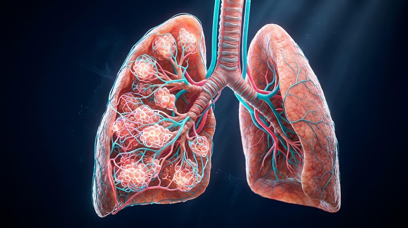
The human respiratory system accomplishes a staggering physiological feat: over the course of a single day, an adult at rest breathes approximately 20,000 times, displacing 10,000 to 12,000 liters of atmospheric air through an interconnected tracheobronchial conduit to facilitate the transfer of over 550 liters of oxygen into systemic circulation. This immense gaseous exchange takes place across the alveolar-capillary membrane�a biological barrier so microscopically thin (averaging just 0.2 to 0.5 micrometers) that its total cumulative surface area exceeds 100 to 130 square meters, roughly equivalent to the size of a regulation singles tennis court, folded into the thoracic cavity.
Understanding pulmonary medicine requires bridging the anatomical continuum from the macro-scale�such as the asymmetric angulation of the primary bronchi and the biomechanical excursion of the thoracic cage�to the sub-microscopic physics of alveolar surface tension, collateral ventilation through the pores of Kohn, and molecular gas diffusion governed by Fick's First Law.
In this clinical masterclass, we explore the 3D anatomical architecture of the tracheobronchial tree, dissect the histological layers of the blood-gas barrier, derive the governing physical equations of gas transport, and examine the pathological destruction seen in chronic obstructive pulmonary disease (COPD) and bronchial asthma using our interactive 3D WebGL anatomical studio.
Interactive 3D Studio: Inspect 3D Pulmonary & Respiratory Tree
Run exact formula simulations on NexProTools.
1. Macroscopic Architecture of the Tracheobronchial Conduit
The conduction zone of the human respiratory tract begins at the larynx and extends through the trachea and 16 successive generations of branching bronchi and bronchioles before reaching the gas-exchanging respiratory zone.
The Trachea and Carinal Bifurcation
The adult human trachea is a midline fibrocartilaginous tube measuring approximately 10 to 12 centimeters in length and 2.0 to 2.5 centimeters in transverse external diameter. Extending from the inferior border of the cricoid cartilage at the level of the sixth cervical vertebra (C6) to the sternal angle of Louis (intervertebral disc space between T4 and T5), the trachea is held continuously patent by 16 to 20 incomplete, anterior C-shaped hyaline cartilage rings.
The posterior gap of each cartilaginous ring is bridged by the fibroelastic trachealis muscle. During the preparatory phase of a vigorous cough, active contraction of the trachealis muscle invaginates the posterior membranous wall into the tracheal lumen, reducing cross-sectional luminal area by up to 60%. According to the fluid dynamic continuity equation:
Where:
Qis volumetric expiratory airflow (\text{m}^3/\text{s})Ais cross-sectional luminal area (\text{m}^2)vis linear flow velocity (\text{m/s})
This luminal narrowing elevates linear air velocities to near-sonic speeds (over 200 to 250 meters per second), generating immense shearing forces capable of dislodging mucus plugs, inhaled particulates, and microbial biofilms from the mucosal surface.
| Anatomical Parameter | Metric Dimension (Adult) | Physiological & Clinical Significance |
|---|---|---|
| Tracheal Length | 10.0 - 12.0 cm | Distance from cricoid cartilage (C6) to carina (T4/T5) |
| Tracheal Internal Diameter | 1.8 - 2.3 cm (Male) / 1.4 - 1.8 cm (Female) | Governs baseline laminar airflow resistance (R \\propto 1/r^4) |
| Hyaline Cartilage Rings | 16 - 20 C-shaped arches | Prevents catastrophic luminal collapse during negative inspiratory pleural pressure |
| Carinal Sub-Bifurcation Angle | 60� - 75� (Normal) / > 90� (Pathologic) | Carinal widening > 90� is a radiographic hallmark of subcarinal adenopathy |
| Right Main Bronchus Length | 2.5 cm (Short & Vertical, ~25� from vertical) | Preferential path for foreign body aspiration and endotracheal hyper-intubation |
| Left Main Bronchus Length | 5.0 cm (Longer, Angled ~45� from vertical) | Traverses beneath the aortic arch and anterior to the descending thoracic aorta |
The 23 Generations of Weibel's Dichotomous Branching
In 1963, Swiss anatomist Ewald Weibel formulated the foundational mathematical model of human lung architecture, establishing that the airway arborizes through 23 symmetrical dichotomous branching generations:
- Conducting Zone (Generations 0 to 16): Trachea, mainstem bronchi, lobar bronchi, segmental bronchi, subsegmental bronchi, down to terminal bronchioles. This zone possesses zero alveoli and constitutes the anatomical dead space (
V_D \\approx 150 \\text{ mL}). Gas transport occurs entirely via bulk convective flow. - Transitional Zone (Generations 17 to 19): Respiratory bronchioles. Alveoli begin to bud intermittently from the bronchiole walls, marking the onset of gas exchange.
- Respiratory Zone (Generations 20 to 23): Alveolar ducts and alveolar sacs. Bulk convection ceases completely, and gas movement occurs entirely via passive molecular diffusion over microscopic distances.
2. Gross Lobar Anatomy & Bronchopulmonary Segments
The human lungs are bilateral, spongy, cone-shaped organs occupying the pleural cavities of the thorax, separated by the central mediastinum.
The Right Lung (Tri-Lobar Architecture)
The right lung is shorter and wider than the left due to the underlying elevation of the right hemidiaphragm by the right lobe of the liver. It has an average adult volume of 3,000 mL and is divided into three distinct lobes by two anatomical fissures:
- The Oblique Fissure: Extends from the posterior chest wall at the T4 level diagonally downward to the sixth costochondral junction, separating the inferior (basal) lobe from the superior and middle lobes.
- The Horizontal (Transverse) Fissure: Arises from the mid-axillary line along the oblique fissure and runs horizontally anteriorly along the fourth costal cartilage, demarcating the superior lobe from the middle lobe.
The right lung contains 10 independent Bronchopulmonary Segments:
- Superior Lobe: Apical (B1), Posterior (B2), Anterior (B3).
- Middle Lobe: Lateral (B4), Medial (B5).
- Inferior Lobe: Superior (B6), Medial Basal (B7), Anterior Basal (B8), Lateral Basal (B9), Posterior Basal (B10).
The Left Lung (Bi-Lobar Architecture & Lingula)
The left lung is slightly smaller (average volume 2,500 mL) to accommodate the asymmetric leftward tilt and apex of the pericardial sac and heart. It possesses only a single oblique fissure dividing it into:
- Superior Lobe: Characterized by the anterior Cardiac Notch (an indentation accommodating the left ventricular apex) and the Lingula (a tongue-like inferior projection corresponding embryologically to the right middle lobe).
- Inferior Lobe: Forms the massive posterior-inferior base of the lung.
The left lung contains 8 to 9 bronchopulmonary segments:
- Superior Lobe: Apicoposterior (B1/B2), Anterior (B3), Superior Lingular (B4), Inferior Lingular (B5).
- Inferior Lobe: Superior (B6), Anteromedial Basal (B7/B8), Lateral Basal (B9), Posterior Basal (B10).
[!NOTE] Each bronchopulmonary segment functions as an autonomous anatomical and surgical unit: it possesses its own dedicated tertiary segmental bronchus, tertiary branch of the pulmonary artery, and autonomous autonomic nervous supply. Pulmonary veins, by contrast, run intersegmentally within the fibrous connective tissue septa separating adjacent segments, facilitating anatomical lung segmentectomy without disrupting neighboring vascular integrity.
3. Microscopic Histology: The Blood-Gas Barrier & Alveolar Acinus
The terminal architectural unit of the lung is the Pulmonary Acinus, defined as the parenchymal region distal to a single terminal bronchiole, measuring approximately 6 to 8 millimeters in diameter and containing roughly 10,000 alveoli.
Cellular Composition of the Alveolus
The adult lungs contain approximately 480 million microscopic alveoli (range: 274 to 790 million), each measuring 200 to 250 micrometers in diameter. The alveolar wall is lined by two distinct epithelial cell types:
1. Type I Pneumocytes (Squamous Alveolar Cells)
Comprise only 8% to 10% of total alveolar cells by number, yet their extreme cytoplasmic attenuation (thinning down to 0.05 to 0.1 micrometers) enables them to cover over 95% of the entire alveolar gas-exchange surface. Type I pneumocytes are terminally differentiated, cannot undergo mitotic division, and are joined to adjacent cells by continuous zonula occludens (tight junctions), forming an impermeable barrier against interstitial fluid transudation into the air spaces.
2. Type II Pneumocytes (Granular Surfactant Synthesizers)
Comprise 15% to 18% of alveolar cells by number but cover less than 5% of surface area. These cuboidal cells contain abundant cytoplasmic organelles and distinctive concentric lamellar bodies packed with Pulmonary Surfactant. Type II cells serve two crucial functions:
- They actively synthesize, secrete, and reuptake pulmonary surfactant.
- They serve as the progenitor stem cells for the alveolar epithelium; following toxic or viral destruction of Type I cells, Type II pneumocytes rapidly proliferate and differentiate into flattened Type I squamous cells.
3. Alveolar Macrophages (Dust Cells)
Patrol the fluid hypophase of the alveolar lumen, phagocytosing inhaled particulates, allergens, silica, and senescent surfactant molecules. They digest bacterial pathogens and migrate upward via the mucociliary escalator toward the pharynx or into lymphatic channels.
4. The Biophysics of Pulmonary Gas Exchange
Gas transfer across the alveolar-capillary membrane is a purely passive physical process driven by partial pressure gradients, surface area, membrane thickness, and molecular solubility.
Fick's First Law of Gas Diffusion
The rate of transfer of a gas across a biological tissue barrier is mathematically formalized by Fick's Law of Diffusion:
Where:
\dot{V}_{\text{gas}}is the volume of gas diffusing per unit time (\text{mL/min})Ais the total cross-sectional surface area of the diffusion barrier (\approx 100\text{ to }130\text{ m}^2)Tis the thickness of the blood-gas barrier (\approx 0.2\text{ to }0.5\text{ }\mu\text{m})(P_1 - P_2)is the trans-membrane partial pressure difference of the gas (\text{mmHg})Dis the diffusion constant of the specific gas, derived from Graham's Law:
Because carbon dioxide (\text{CO}_2) is approximately 24 times more soluble in aqueous biological membranes than oxygen (\text{O}_2), the diffusion constant for \text{CO}_2 is roughly 20 times greater than that of \text{O}_2:
Consequently, in pathological states characterized by alveolar membrane thickening (such as idiopathic pulmonary fibrosis or acute respiratory distress syndrome), hypoxemia (P_{a\text{O}_2} < 60\text{ mmHg}) occurs long before hypercapnia (P_{a\text{CO}_2} > 45\text{ mmHg}).
The Alveolar Gas Equation
To calculate the ideal alveolar oxygen tension (P_{A\text{O}_2}) available for capillary equilibration at sea level:
Under standard physiological conditions:
P_{\text{atm}} = 760\text{ mmHg}(barometric pressure at sea level)P_{\text{H}_2\text{O}} = 47\text{ mmHg}(water vapor pressure at body temperature 37�C)F_i\text{O}_2 = 0.21(fraction of inspired oxygen in ambient room air)P_{a\text{CO}_2} = 40\text{ mmHg}(arterial carbon dioxide tension)R = 0.8(respiratory exchange ratio:\dot{V}_{\text{CO}_2} / \dot{V}_{\text{O}_2})
Laplace's Law and the Crucial Role of Surfactant
An alveolus can be idealized as a sphere open to atmospheric pressure through its conducting bronchiole. The inward collapsing pressure (P) generated by the liquid air-water surface tension (\gamma) on the alveolar wall is governed by the Law of Young-Laplace:
Where:
Pis collapsing pressure (\text{dynes/cm}^2or\text{cmH}_2\text{O})\gammais surface tension (\text{dynes/cm})ris alveolar radius (\text{cm})
Surfactant is composed of 80% phospholipids (predominantly dipalmitoylphosphatidylcholine [DPPC]), 10% neutral lipids, and 10% surfactant-associated proteins (SP-A, SP-B, SP-C, and SP-D). The hydrophobic fatty acyl tails of DPPC project outward into the alveolar gas space, while the hydrophilic polar head groups orient downward into the aqueous hypophase, disrupting the strong intermolecular hydrogen bonds between water molecules.
As alveoli deflate during expiration (r decreases), DPPC molecules are compressed tightly together, driving surface tension down from a baseline of 50\text{ dynes/cm} to near 1\text{ to }5\text{ dynes/cm}, eliminating collapsing pressure and preventing end-expiratory microatelectasis.
5. Clinical Pathophysiology: COPD vs. Bronchial Asthma
Obstructive lung diseases represent one of the leading global causes of morbidity and mortality, characterized by limitation of expiratory airflow that is not fully reversible (COPD) or variable and reversible (Asthma).
| Clinical Parameter | Chronic Obstructive Pulmonary Disease (COPD) | Bronchial Asthma |
|---|---|---|
| Primary Etiology | Chronic cigarette smoke / biomass fuel exposure | Genetic atopy + environmental allergen sensitization |
| Typical Age of Onset | > 40 - 45 years of age | Childhood or early adulthood (< 25 years) |
| Airflow Reversibility | Largely irreversible (post-bronchodilator \text{FEV}_1/\text{FVC} < 0.70) | Highly reversible (\Delta\text{FEV}_1 > 12\% and > 200\text{ mL}) |
| Dominant Inflammatory Cells | CD8+ T-lymphocytes, Neutrophils, Macrophages | Eosinophils, CD4+ Th2 lymphocytes, Mast cells, ILC2 |
| Primary Cytokine Mediators | TNF-\alpha, IL-8, Leukotriene B4, MMP-9, MMP-12 | IL-4, IL-5, IL-13, Histamine, Leukotriene C4/D4 |
| Structural Pathology | Alveolar septal destruction (Emphysema) + Mucus metaplasia | Smooth muscle hypertrophy + Basement membrane thickening |
Diffusing Capacity (\text{DLCO}) | Markedly reduced (< 60\text{ - }70\% predicted) | Normal or mildly elevated (> 100\% predicted) |
| Response to Inhaled Steroids | Variable / modest; reduces exacerbation frequency | First-line foundational controller therapy |
Chronic Obstructive Pulmonary Disease (COPD)
COPD is an inflammatory syndrome encompassing two major pathological phenotypes that frequently coexist in varying proportions:
1. Pulmonary Emphysema
Defined histopathologically as permanent, abnormal enlargement of airspaces distal to terminal bronchioles accompanied by destructive lysis of alveolar septal walls without obvious fibrosis.
- Pathogenetic Mechanism: Chronic exposure to tobacco smoke activates alveolar macrophages and recruits polymorphonuclear neutrophils, which release massive quantities of serine proteinases, particularly neutrophil elastase (NE) and matrix metalloproteinases (MMP-9, MMP-12). In healthy lungs, these proteinases are inhibited by endogenous alpha-1 antitrypsin (AAT). In smokers, cigarette smoke oxidizes methionine-358 at the active site of AAT, rendering it inactive and triggering an unstoppable protease-antiprotease imbalance that digests elastin and collagen scaffolding.
- Biomechanical Consequence: Destruction of alveolar attachments leads to loss of lung elastic recoil. Because radial tethering traction is lost, small non-cartilaginous airways dynamically collapse during forced expiration, trapping gas within the acini and producing severe static and dynamic hyperinflation (elevated Residual Volume [RV] and Functional Residual Capacity [FRC]).
2. Chronic Bronchitis
Defined clinically as a persistent cough with sputum production for at least 3 consecutive months in 2 successive years. Histologically characterized by:
- Hypertrophy and hyperplasia of submucosal mucus glands (elevated Reid Index
> 0.50, where the Reid index is the ratio of gland layer thickness to total bronchial wall thickness). - Squamous metaplasia of pseudostratified ciliated columnar respiratory epithelium, paralyzing mucociliary clearance.
- Luminal obstruction by thick, viscous mucus plugs harboring bacterial colonizers (Haemophilus influenzae, Streptococcus pneumoniae, Moraxella catarrhalis).
Bronchial Asthma Biomechanics & Airway Remodeling
Asthma is a chronic inflammatory disorder of the conducting airways driven primarily by a Type 2 helper T-cell (Th2) immune phenotype:
- Sensitization & IgE Cross-Linking: Inhaled allergens (dust mites, animal dander, pollen) are captured by dendritic cells and presented to na�ve T-cells, driving Th2 differentiation. Th2 cells secrete interleukin-4 (IL-4) and IL-13, stimulating B-cell class switching to immunoglobulin E (IgE).
- Mast Cell Deegranulation: IgE binds to high-affinity Fc
\varepsilonRI receptors on tissue mast cells. Allergen re-exposure cross-links bound IgE, causing immediate release of preformed histamine, prostaglandin D2, and cysteinyl leukotrienes (LTC_4, LTD_4, LTE_4). - Acute Bronchospasm: Leukotrienes bind to CysLT1 receptors on bronchial smooth muscle cells, triggering an intense, rapid bronchoconstriction that increases airway resistance inversely proportional to the fourth power of the radius according to Poiseuille's law:
Where:
\etais dynamic viscosity of airLis airway lengthris internal airway radius
A mere 50% reduction in airway luminal radius results in a 16-fold (2^4 = 16) increase in total airway resistance, producing audible expiratory wheezing, tachypnea, and prolonged expiratory phase.
- Chronic Airway Remodeling: Repeated cycles of allergic inflammation induce irreversible architectural remodeling:
- Markedly thickened reticular basement membrane (subepithelial collagen deposition).
- Massive hyperplasia and hypertrophy of airway smooth muscle bundles.
- Angiogenic proliferation of subepithelial microvessels.
- Goblet cell hyperplasia with extensive intra-luminal mucin-5AC hypersecretion.
6. Diagnostic Spirometry & Quantitative Pulmonary Function
Diagnostic evaluation of respiratory symptoms relies upon standardized spirometry and plethysmographic lung volume determinations performed in accordance with American Thoracic Society / European Respiratory Society (ATS/ERS) standards.
| Spirometric Index | Normal Reference Value | COPD Pattern | Asthma Pattern |
|---|---|---|---|
\text{FEV}_1 (Forced Expiratory Volume in 1 sec) | \ge 80\% predicted | Progressively reduced (< 80\%) | Reduced during attack; normal in remission |
\text{FVC} (Forced Vital Capacity) | \ge 80\% predicted | Normal or reduced (air trapping) | Normal or mildly reduced |
\text{FEV}_1/\text{FVC} Ratio | 0.70\text{ - }0.80 (> \text{LLN}) | < 0.70 (Fixed Obstruction) | < 0.70 during bronchospasm; normalizes with SABA |
| Bronchodilator Reversibility | < 12\% and < 200\text{ mL} | Negative or partial (< 12\%) | Positive (\ge 12\% and \ge 200\text{ mL} increase) |
Total Lung Capacity (\text{TLC}) | 80\text{ - }120\% predicted | Elevated (> 120\%, Hyperinflation) | Normal or mildly elevated |
Residual Volume (\text{RV}) | 80\text{ - }120\% predicted | Markedly elevated (> 140\%, Air Trapping) | Mildly elevated during acute exacerbations |
\text{DLCO} (Gas Diffusion) | 80\text{ - }120\% predicted | Markedly reduced (< 60\%) | Normal or elevated (> 100\%) |
The Global Initiative for Chronic Obstructive Lung Disease (GOLD) Staging
In patients with a confirmed post-bronchodilator \text{FEV}_1/\text{FVC} < 0.70, airflow limitation is classified into 4 severity grades based on \text{FEV}_1:
- GOLD 1 (Mild):
\text{FEV}_1 \ge 80\%predicted - GOLD 2 (Moderate):
50\% \le \text{FEV}_1 < 80\%predicted - GOLD 3 (Severe):
30\% \le \text{FEV}_1 < 50\%predicted - GOLD 4 (Very Severe):
\text{FEV}_1 < 30\%predicted
Global Initiative for Asthma (GINA) Track 1 Management
Modern asthma guidelines emphasize the immediate avoidance of short-acting beta-2 agonist (SABA) monotherapy due to increased risk of fatal exacerbations and downregulation of beta-2 receptors. Instead, GINA Track 1 mandates an Inhaled Corticosteroid (ICS) - Formoterol combination as the foundational reliever across all severity steps:
- Step 1 - 2: As-needed low-dose ICS-formoterol for symptom relief.
- Step 3: Low-dose maintenance and reliever ICS-formoterol.
- Step 4: Medium-dose maintenance and reliever ICS-formoterol.
- Step 5: High-dose maintenance ICS-formoterol + add-on long-acting muscarinic antagonist (LAMA, e.g., Tiotropium) or targeted biologic therapies:
- Anti-IgE (Omalizumab) for severe allergic asthma with elevated serum IgE.
- Anti-IL-5 (Mepolizumab, Reslizumab) or Anti-IL-5R
\alpha(Benralizumab) for severe eosinophilic asthma. - Anti-IL-4R
\alpha(Dupilumab) blocking both IL-4 and IL-13 signaling. - Anti-TSLP (Tezepelumab) targeting upstream thymic stromal lymphopoietin.
7. Laboratory Biomarkers & ICD-10 Coding Reference
Comprehensive respiratory evaluation integrates physiological testing with specific molecular biomarkers:
| Biomarker Assay | Standard Physiological Range | Clinical Significance in Respiratory Disease |
|---|---|---|
| Alpha-1 Antitrypsin (AAT) | 100 - 220\text{ mg/dL} (20 - 53\text{ }\mu\text{mol/L}) | Levels < 57\text{ mg/dL} indicative of severe deficiency (PiZZ genotype), driving premature basal emphysema. |
| Fractional Exhaled Nitric Oxide (FeNO) | < 25\text{ ppb} (Normal) / > 50\text{ ppb} (High) | Directly reflects IL-4/IL-13 induced inducible nitric oxide synthase (iNOS) activity; predicts corticosteroid responsiveness. |
| Blood Eosinophil Count | < 150\text{ cells/}\mu\text{L} (Low) / \ge 300\text{ cells/}\mu\text{L} (High) | Identifies Type 2 inflammation in COPD; predicts substantial benefit from adding inhaled corticosteroids to dual bronchodilators. |
Arterial Blood Gas: P_{a\text{O}_2} | 80 - 100\text{ mmHg} on room air | Resting P_{a\text{O}_2} \le 55\text{ mmHg} (or \le 59\text{ mmHg} with cor pulmonale) qualifies for long-term home oxygen therapy. |
Arterial Blood Gas: P_{a\text{CO}_2} | 35 - 45\text{ mmHg} | Elevated P_{a\text{CO}_2} > 45\text{ mmHg} indicates alveolar hypoventilation, respiratory fatigue, or severe ventilation-perfusion mismatch. |
| Serum Total IgE | < 100\text{ kU/L} | Screening metric for allergic sensitization; determines dosing thresholds for anti-IgE biologic therapy (Omalizumab). |
ICD-10 Diagnosis Codes for Pulmonary Conditions
- J44.0: Chronic obstructive pulmonary disease with acute lower respiratory infection
- J44.1: Chronic obstructive pulmonary disease with acute exacerbation, unspecified
- J44.9: Chronic obstructive pulmonary disease, unspecified
- J43.9: Emphysema, unspecified
- J45.20: Mild intermittent asthma, uncomplicated
- J45.41: Moderate persistent asthma with acute exacerbation
- J45.52: Severe persistent asthma with status asthmaticus
- E88.01: Alpha-1-antitrypsin deficiency
- J84.112: Idiopathic pulmonary fibrosis
8. Summary & Interactive 3D Exploration
The respiratory system represents an elegant balance of mechanical compliance, aerodynamic branching geometry, and molecular diffusion physics. By combining macroscopic knowledge of lobar and carinal morphology with microstructural appreciation of surfactant physics, clinicians can accurately diagnose and target the underlying drivers of obstructive, restrictive, and vascular lung disease.
Use our Interactive 3D Anatomy & Clinical Studio to manipulate the 3D tracheobronchial arborization, examine the terminal alveolar sac clusters, toggle dynamic respiratory ventilation, and simulate the parenchymal destruction of pulmonary emphysema in real-time.