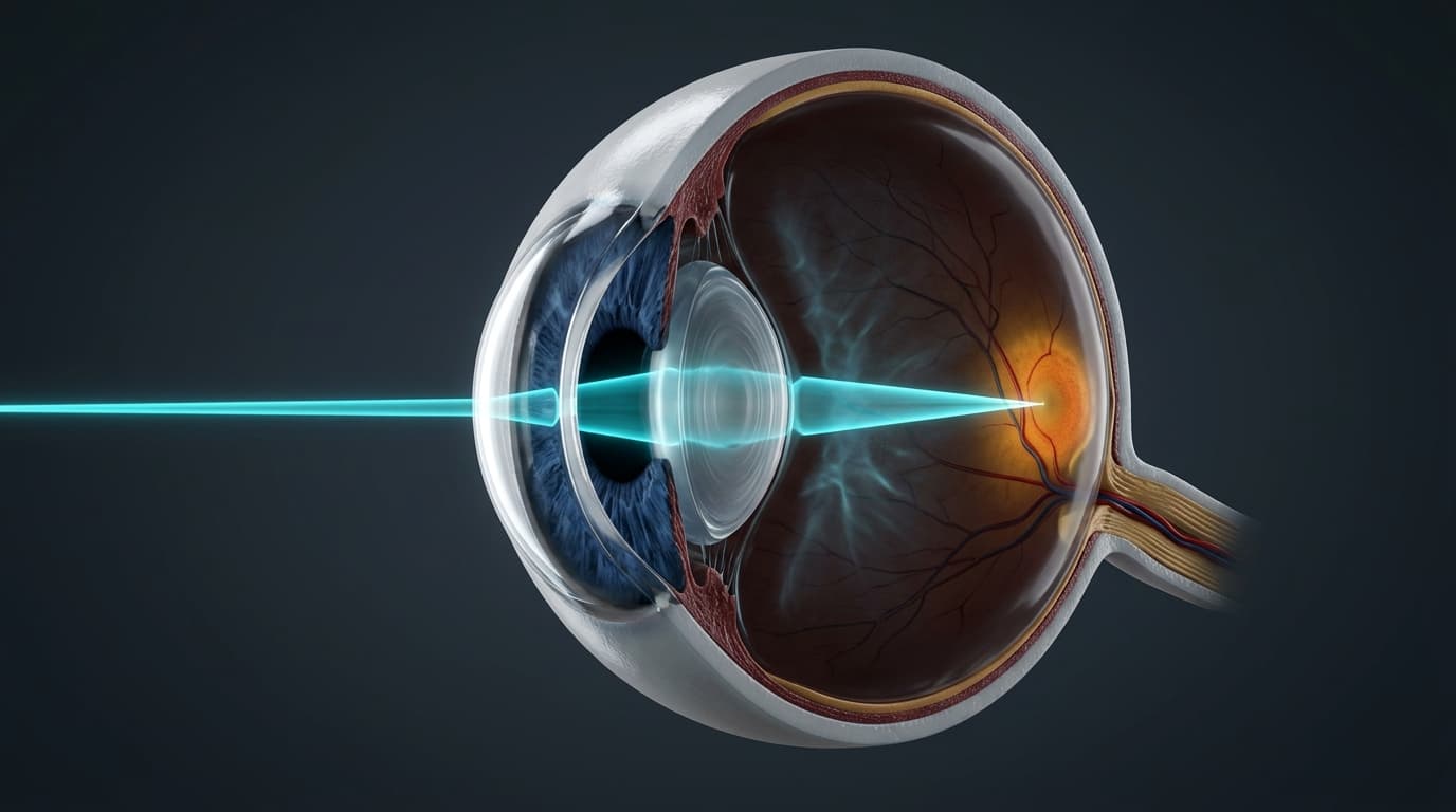
The human eye is one of nature's most sophisticated optical imaging instruments, transforming ambient photons of the visible electromagnetic spectrum (wavelengths roughly 380 to 740 nanometers) into spatial arrays of electrical action potentials interpreted by the visual cortex. Measuring on average just 24 millimeters in axial anteroposterior diameter in the emmetropic adult, the ocular globe coordinates a two-element compound refractive lens system�comprising the anterior corneal dome and the dynamic, biconvex crystalline lens�to focus parallel incoming light rays precisely onto a microscopic 1.5-millimeter photoreceptor pit known as the fovea centralis.
Understanding ophthalmic pathology requires analyzing the eye from both a structural macroscopic perspective�such as the three concentric ocular tunics and the aqueous fluid drainage pathways�and a biophysical perspective governing dioptric refraction, Snell's law, Helmholtz accommodation mechanics, and retinal ganglion cell neuroprotection.
In this clinical masterclass, we explore the high-resolution 3D anatomy of the human eye, derive the mathematical optical principles of ocular refraction, dissect the fluid mechanics of aqueous humor outflow, and examine the cellular mechanisms of axial myopia, cataracts, and glaucomatous optic neuropathy using our interactive 3D WebGL anatomical workstation.
Interactive 3D Studio: Inspect 3D Eye & Visual Optics
Run exact formula simulations on NexProTools.
1. Macroscopic Architecture of the Ocular Globe
The adult human eyeball resides within the bony quadrilateral orbital cavity, cushioned by retrobulbar adipose tissue and suspended by six extraocular muscles and Tenon's capsule. The wall of the globe is composed of three concentric architectural tunics:
| Ocular Metric | Normal Emmetropic Value | Clinical & Diagnostic Significance |
|---|---|---|
| Axial Length (AL) | 23.5 - 24.5 mm | An increase of just 1.0 mm produces approximately -2.5 to -3.0 Diopters of axial myopia |
| Total Ocular Refractive Power | +58.0 to +60.0 Diopters | Optical power required to focus infinite rays onto the retina |
| Corneal Refractive Power | +43.0 to +44.0 Diopters | Contributes roughly 70% to 75% of total static ocular refraction |
| Crystalline Lens Power (Rest) | +15.0 to +20.0 Diopters | Dynamic lens providing variable accommodation for near vision |
| Central Corneal Thickness (CCT) | 535 - 550 �m | Thin CCT (< 500 �m) leads to artificial underestimation of Goldman IOP |
| Anterior Chamber Depth (ACD) | 3.0 - 3.4 mm | Shallow ACD (< 2.5 mm) is a primary predisposing risk factor for angle-closure glaucoma |
| Intraocular Pressure (IOP) | 10 - 21 mmHg | Equilibrium between ciliary body aqueous secretion and trabecular outflow |
2. Geometric Optics of the Eye: Corneal Refraction & Snell's Law
Refraction occurs whenever an electromagnetic wave traverses an optical boundary separating two media of differing optical densities (refractive indices). The physical angle of deviation is governed by Snell's Law of Refraction:
Where:
n_1is the refractive index of the incident medium\theta_1is the angle of incidence relative to the surface normaln_2is the refractive index of the transmitting medium\theta_2is the angle of refracted transmission
Because the difference in refractive index across the air-corneal interface (\Delta n = 1.376 - 1.000 = 0.376) is dramatically larger than across the aqueous-lens interface (\Delta n = 1.406 - 1.336 = 0.070), the anterior surface of the cornea accounts for more than two-thirds (+43 to +44 Diopters) of the entire eye's focusing power.
The Dioptric Power Equation for Spherical Surfaces
The optical vergence power (D, measured in Diopters, \text{m}^{-1}) of a curved spherical refracting boundary is calculated using the single-surface power formula:
Where:
Pis refractive power in Dioptersn_2 - n_1is the difference in refractive indexRis the radius of curvature of the anterior optical surface in meters
For an anterior corneal radius of curvature R = 7.7\text{ mm} = 0.0077\text{ m}:
When accounting for the divergent negative power (-5.8 D) of the posterior corneal-aqueous interface (R = 6.8\text{ mm}), the net static refractive power of the human cornea stabilizes at +43.05 Diopters.
3. The Crystalline Lens & Biomechanics of Accommodation
While the cornea provides fixed, static refraction, the biconvex crystalline lens provides dynamic, adjustable focusing power through the physiological process of Accommodation, enabling the eye to bring near objects into sharp focus.
The Helmholtz Theory of Accommodation
In 1855, German polymath Hermann von Helmholtz formulated the classic mechanism of accommodation:
- Distant Vision (Unaccommodated State): The circular ciliary muscle fibers are fully relaxed, expanding the ciliary ring diameter. This exerts continuous centrifugal tensile stress on the suspensory Zonules of Zinn, pulling outward on the elastic lens capsule. The crystalline lens is stretched into a flattened, thinned biconvex shape with an optical power of approximately +15 to +18 Diopters.
- Near Vision (Accommodated State): Parasympathetic cholinergic signaling via the Edinger-Westphal nucleus and Cranial Nerve III stimulates contraction of the ciliary muscle. As the muscle fibers contract toward the lens equator, the diameter of the ciliary ring constricts.
- Zonular Relaxation & Elastic Recoil: The tension on the zonular fibers drops to near zero. Released from external stretching, the inherent elasticity of the crystalline lens capsule forces the pliable lens substance into a more steeply curved, thicker biconvex profile. The anterior radius of curvature steepens from
10.0\text{ mm}down to5.5\text{ mm}, increasing optical power by up to +8 to +12 Diopters.
The Pathophysiology of Presbyopia
Accommodation is not lost due to weakness of the ciliary muscle; rather, it is caused by the progressive, lifelong accumulation of lens fibers:
- The lens epithelium continuously replicates, laying down new secondary lens fibers that compress the central lens nucleus.
- As lens proteins (alpha, beta, and gamma crystallins) undergo oxidative cross-linking and insoluble aggregation, the lens substance hardens (Nuclear Sclerosis).
- Concurrently, the basement membrane lens capsule loses its youthful elastic recoil.
- By age 45 to 50, the maximal amplitude of accommodation drops from
14\text{ Diopters}(at age 10) to< 2\text{ Diopters}, requiring reading glasses (+1.50 to +2.50 D convex plus lenses) to substitute for lost intrinsic vergence.
4. Retinal Histology & The Phototransduction Cascade
The retina is a specialized, ten-layered extension of the central nervous system lining the posterior two-thirds of the globe.
The Molecular Cascade of Phototransduction
Phototransduction is counterintuitive compared to conventional sensory receptors: in the dark, photoreceptors are continuously depolarized (-40 mV) and tonically release glutamate; light absorption causes hyperpolarization (-70 mV), shutting off neurotransmitter release:
- Photon Absorption: A photon strikes the photopigment Rhodopsin (embedded in outer segment membranous discs), causing photoisomerization of the chromophore 11-cis-retinal into all-trans-retinal.
- Conformational Activation: This geometric isomerization induces a conformational shift into active Metarhodopsin II.
- G-Protein Transducin Coupling: Metarhodopsin II binds and activates hundreds of molecules of the heterotrimeric G-protein Transducin (
G_t), catalyzing GTP exchange on the alpha subunit (G_{\alpha t}). - PDE6 Activation: Active
G_{\alpha t}\text{-GTP}complexes dissociate and bind the inhibitory gamma subunits of cGMP Phosphodiesterase 6 (PDE6), unleashing catalytic PDE6 activity. - cGMP Hydrolysis: PDE6 hydrolyzes cyclic GMP into 5'-GMP at immense catalytic velocities (over 2,000 cGMP molecules hydrolyzed per second per activated rhodopsin).
- Cation Channel Closure: The drop in cytoplasmic cGMP concentration causes cGMP to dissociate from cyclic nucleotide-gated (CNG) cation channels in the plasma membrane.
- Hyperpolarization: Inward sodium and calcium currents (
I_{\text{dark}}) cease, while potassium continues to leak outward through inner segment channels. The membrane potential hyperpolarizes from-40\text{ mV}to-70\text{ mV}, shutting off voltage-gated calcium channels and stopping glutamate exocytosis into the outer plexiform layer.
5. Refractive Errors: Myopia, Hyperopia & Astigmatism
Refractive errors occur whenever the optical focal point of the relaxed eye fails to coincide precisely with the neurosensory retinal plane.
| Refractive Anomaly | Structural Mechanism | Focal Point Relative to Retina | Optical Lens Correction |
|---|---|---|---|
| Emmetropia (Normal) | Axial length (24 mm) matches +60 D focal length | Precisely at foveal photoreceptor plane | None required (Plano) |
| Axial Myopia (Nearsighted) | Globe elongation ($> 24.5\text$) | In front of retina (in vitreous body) | Concave Minus (Biconcave) Lens |
| Hyperopia (Farsighted) | Globe short ($< 22.5\text$) or flat cornea | Behind the anatomical plane of the retina | Convex Plus (Biconvex) Lens |
| Regular Astigmatism | Asymmetric corneal curvature (toric surface) | Two distinct focal lines at 90� to each other | Cylindrical / Toric Lens |
| Presbyopia | Sclerosis of crystalline lens core | Near point recedes beyond working distance | Convex Plus Reading Addition (+Add) |
Axial Myopia Biomechanics
In axial myopia, excessive post-natal elongation of the scleral shell causes parallel light rays from a distant object to converge into a focal point in the mid-vitreous body, diverging again before striking the retina as a blurred blur circle:
- Every 1 millimeter increase in axial length produces approximately -2.70 Diopters of myopia.
- Pathological high myopia (axial length $> 26.5\text$ or sphere $< -6.00\text$) stretches the sclera, choroid, and retina, creating severe structural vulnerabilities:
- Posterior Staphyloma: Ectatic outpouching of the thinned posterior scleral wall.
- Lattice Degeneration & Retinal Detachment: Peripheral retinal thinning leading to atrophic holes, horse-shoe tears, and rhegmatogenous retinal detachment.
- Myopic Maculopathy: Bruch's membrane lacquer cracks and choroidal neovascularization (CNV).
When total optical power is +60 D, the ideal focal distance behind the principal plane is 16.7 mm (total axial length ~24 mm). If axial length stretches to 26 mm, the focal point lands 2.0 mm anterior to the photoreceptors, requiring a divergent minus lens to push the focal point backward.
6. Aqueous Humor Dynamics & Pathophysiology of Glaucoma
Glaucoma is a group of progressive optic neuropathies characterized by apoptosis of retinal ganglion cell axons, excavated cupping of the optic nerve head, and visual field loss. Elevated intraocular pressure (IOP) is the single most important modifiable risk factor.
The Goldmann Equation for Intraocular Pressure
Steady-state intraocular pressure is mathematically modeled by the Goldmann Equation:
Where:
P_{\text{IOP}}is intraocular pressure (mmHg)Fis rate of aqueous humor formation by ciliary processes (\approx 2.5\ \mu\text{L/min})Uis uveoscleral outflow rate (\approx 0.25\ \mu\text{L/min})Cis facility of trabecular outflow (\approx 0.28\ \mu\text{L/min/mmHg})P_vis episcleral venous pressure (\approx 8\text{ to }10\text{ mmHg})
In Primary Open-Angle Glaucoma (POAG), extracellular matrix glycosaminoglycans, cross-linked actin networks, and myocilin aggregates accumulate within the Juxtacanalicular Trabecular Meshwork, dramatically reducing outflow facility C. As C plummets, P_{\text{IOP}} rises from normal (14\text{ mmHg}) into pathological ranges (24\text{ to }40\text{ mmHg}).
Mechanism of Glaucomatous Axonal Death
High IOP exerts continuous mechanical strain on the fenestrated Lamina Cribrosa at the posterior scleral canal:
- Lamina cribrosa plates deflect backward, compressing traversing unmyelinated retinal ganglion cell (RGC) axons.
- Orthograde and retrograde axoplasmic transport of neurotrophins (Brain-Derived Neurotrophic Factor, BDNF) is blocked at the cribriform pores.
- Deprived of target-derived trophic support, RGCs initiate caspase-dependent apoptotic cell death.
- Concurrently, microvascular ischemia of the optic nerve head occurs when IOP exceeds capillary perfusion pressure.
- The neuroretinal rim thins progressively, manifesting clinically as an enlarged Cup-to-Disc (C/D) Ratio ($> 0.6$ or asymmetric $> 0.2$).
7. Diagnostic Testing Reference & ICD-10 Classification
Accurate ophthalmological evaluation integrates structural tomography with functional psychophysical visual field assessment.
| Diagnostic Modality | Normal Physiological Reference | Clinical Significance in Ocular Pathology |
|---|---|---|
| Goldmann Applanation Tonometry (IOP) | $10 - 21\text$ | Standard of care; requires CCT correction for true corneal biomechanics |
| Optical Coherence Tomography (RNFL) | $85 - 110\text\mu\text$ (Average thickness) | Sub-micron cross-sectional imaging measuring peripapillary retinal nerve fiber layer loss |
| Humphrey Visual Field (24-2 SITA-Fast) | Mean Deviation (MD) $> -2.00\text$ | Detects early glaucomatous nasal steps, arcuate Bjerrum scotomas, and paracentral deficits |
| Endothelial Specular Microscopy | $2,000 - 3,000\text^2$ | Evaluates corneal endothelial pump reserve before intraocular cataract surgery |
| Axial Biometry (IOLMaster / Lenstar) | $22.0 - 24.5\text$ | Determines intraocular lens power calculation (Barrett Universal II / Hill-RBF formulas) |
ICD-10 Diagnosis Codes for Ocular Conditions
- H52.13: Myopia, bilateral
- H52.03: Hypermetropia, bilateral
- H52.203: Unspecified astigmatism, bilateral
- H52.4: Presbyopia
- H40.1130: Primary open-angle glaucoma, bilateral, unspecified stage
- H40.223: Chronic angle-closure glaucoma, bilateral
- H25.13: Age-related nuclear cataract, bilateral
- H35.31: Nonexudative age-related macular degeneration (Dry AMD)
- H35.32: Exudative age-related macular degeneration (Wet AMD)
8. Summary & Interactive 3D Visual Optics Studio
The human eye exemplifies the seamless integration of dioptric geometric optics and advanced neurobiology. From the steep +43 D refractive power of the anterior corneal dome to the nanometer-scale precision of the foveal cone mosaic, understanding ocular mechanics unlocks deep clinical insights into laser vision correction (LASIK/PRK), premium intraocular lens selection, and neuroprotective glaucoma therapies.
Explore our Interactive 3D Human Anatomy Studio to rotate the full ocular globe, inspect the internal crystalline lens and ciliary body, trace simulated light ray paths, and activate pathology mode to observe the anatomical focal shifts of myopia and cataract opacification in real-time.