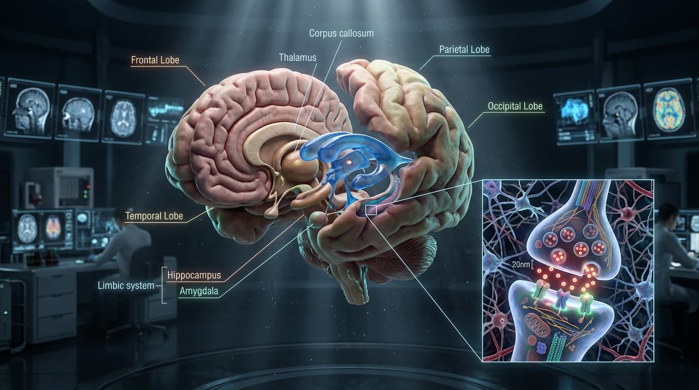
The human brain is the most geometrically complex and energetically demanding structure in the known universe. Weighing approximately 1.4 kilograms�representing just 2% of total body mass�it consumes over 20% of the body's total basal oxygen and glucose supply. Within its convoluted cortical mantle reside roughly 86 billion neurons, interconnected by more than 100 trillion synaptic junctions operating across a spatial scale ranging from centimeters of cerebral white matter tracts down to twenty nanometers of fluid gap across the synaptic cleft.
To comprehend modern clinical neurology, cognitive neuroscience, and neuropharmacology, one must be able to bridge these dimensional realms: understanding how the macroscopic topography of cerebral lobes coordinates with the deep limbic circuits of memory consolidation, how the ventricular glymphatic network flushes metabolic waste during slow-wave sleep, and how neurotransmitter vesicles release their chemical cargo to bind postsynaptic transmembrane receptors.
In this clinical masterclass, we explore the articulated 3D anatomy of the human brain, trace the functional mapping of cerebral lobes, examine the ventricular cerebrospinal fluid pathways, derive the kinetics of synaptic transmission, and simulate competitive drug receptor blockade using our interactive 3D WebGL workstation.
Interactive 3D Studio: Explore 3D Brain Lobes & Limbic Core
Run exact formula simulations on NexProTools.
1. Telencephalic Topography: The Four Cerebral Lobes & Functional Cortical Mapping
The cerebral cortex is organized into two symmetrical cerebral hemispheres separated by the deep Longitudinal Cerebral Fissure and interconnected by the massive, 200-million-axon commissural highway of the Corpus Callosum.
The convoluted folding of the cortex into Gyri (crests) and Sulci (grooves) expands the total cortical surface area to roughly 2,200 cm��more than two-thirds of which lies hidden within the depths of the sulci.
| Cerebral Lobe | Key Structural Boundaries | Primary Cortical Specializations | Clinical Deficit Upon Injury | | :--- | :--- | :--- | :--- | | **Frontal Lobe** | Anterior to Central Sulcus of Rolando | Motor execution (Area 4), working memory, executive cognitive control, expressive speech (Broca's) | Contralateral paresis, Broca's motor aphasia, abulia, behavioral disinhibition | | **Parietal Lobe** | Between Central and Parieto-Occipital sulci | Primary somatosensory perception (Areas 3, 1, 2), body scheme, visuomotor integration | Astereognosis, hemispatial neglect (non-dominant), Gerstmann syndrome | | **Temporal Lobe** | Inferior to Lateral Sylvian Fissure | Primary audition (A1), receptive language (Wernicke's), declarative memory encoding | Wernicke's fluent aphasia, anterograde amnesia, auditory agnosia | | **Occipital Lobe** | Posterior to Parieto-Occipital notch | Primary visual cortex (Striate V1, Area 17), retinotopic spatial mapping, motion and form | Homonymous hemianopia, cortical blindness, visual agnosia |
1. The Frontal Lobe & Motor Homunculus
Extending from the frontal pole to the Central Sulcus of Rolando:
- Primary Motor Cortex (Brodmann Area 4): Located in the precentral gyrus. It contains the giant pyramidal Betz cells whose axons descend through the internal capsule to form the corticospinal tract. It displays an inverted topographical map of the contralateral body�the Motor Homunculus�with disproportionately massive representation dedicated to the hands, fingers, lips, and tongue (reflecting evolutionary manual dexterity and vocal articulation).
- Prefrontal Cortex (
PFC): Comprising the dorsolateral (dlPFC), orbitofrontal (OFC), and ventromedial (vmPFC) regions. It serves as the brain's executive conductor: maintaining goal-directed working memory, inhibiting impulsive behavioral reflexes, weighing probabilistic outcomes, and executing abstract rule-based reasoning. - Broca's Expressive Speech Area (Brodmann Areas 44 and 45): Located in the pars opercularis and pars triangularis of the inferior frontal gyrus (dominant left hemisphere in 95% of right-handed and 70% of left-handed individuals). Lesions produce Broca's Motor Aphasia: non-fluent, effortful, telegraphic speech with preserved auditory comprehension.
2. The Parietal Lobe & Somatosensory Representation
- Primary Somatosensory Cortex (Brodmann Areas 3, 1, and 2): Located in the postcentral gyrus. Receives thalamocortical sensory projections from the ventral posterolateral (
VPL) and ventral posteromedial (VPM) nuclei, encoding contralateral discriminative touch, vibration, joint position proprioception, and temperature. - Superior Parietal Lobule: Coordinates body spatial orientation and mental rotation.
- Inferior Parietal Lobule (Supramarginal & Angular Gyri): Integrates multimodal sensory processing. Damage to the non-dominant (right) parietal lobe produces dramatic Hemispatial Neglect Syndrome, where patients completely ignore the left half of their sensory world, failing to dress the left side of their body or eat food from the left half of a plate.
3. The Temporal Lobe: Memory, Language & Audition
- Primary Auditory Cortex (Heschl's Gyri, Brodmann Areas 41 and 42): Tonotopically organized to process auditory sound frequencies.
- Wernicke's Receptive Language Area (Brodmann Area 22): Located in the posterior superior temporal gyrus. Lesions produce Wernicke's Fluent Aphasia: effortless, melodious, grammatically fluent speech that is completely devoid of semantic meaning ("word salad"), accompanied by profound deficits in auditory language comprehension.
- Medial Temporal Lobe: Encapsulates the parahippocampal gyrus, entorhinal cortex, and the hippocampal formation.
4. The Occipital Lobe & Dual Visual Processing Streams
- Primary Visual Cortex (
V1/ Calcarine Cortex, Brodmann Area 17): Retinotopically maps the contralateral visual hemifield. - Information exits
V1along two divergent visual processing highways:- The Dorsal "Where / How" Stream: Projects into the parietal cortex, processing spatial motion, object location, and visual guidance of motor grasping.
- The Ventral "What" Stream: Projects into the inferior temporal cortex (fusiform gyrus), processing high-resolution object recognition, color constancy, and facial identification. Damage to the fusiform face area causes Prosopagnosia (inability to recognize familiar human faces).
2. The Cerebellum & Brainstem: Precision Coordination & Autonomic Control
Positioned beneath the tentorium cerebelli in the posterior cranial fossa, the cerebellum and brainstem operate as the subcortical regulatory foundation of human motor and autonomic physiology.
| Anatomical Sub-Division | Key Nuclei & Fiber Tracts | Physiological Motor / Autonomic Function | | :--- | :--- | :--- | | **Midbrain (Mesencephalon)** | Substantia Nigra (SNc), Red Nucleus, Superior/Inferior Colliculi | Dopaminergic motor modulation, visual tracking reflexes, and auditory startle responses | | **Pons (Metencephalon)** | Transverse Pontine Fibers, Locus Coeruleus, Pneumotaxic Center | Cortico-cerebellar relay bridge, central noradrenaline generation, respiratory rate regulation | | **Medulla Oblongata (Myelencephalon)** | Pyramidal Decussation, Inferior Olives, Pre-B�tzinger Complex | Contralateral motor tract crossing, baroreceptor cardiovascular regulation, autonomic respiratory drive | | **Cerebellar Hemispheres** | Purkinje Cell Layer, Dentate Nucleus, Granule Cell Folia | Predictive motor error correction, balance maintenance, saccadic gaze coordination |
The Cerebellum: 50% of the Brain's Total Neurons
Although the cerebellum accounts for only 10% of total brain volume, its tightly packed horizontal Folia contain more than 50 billion neurons�over half the total neuronal population of the entire central nervous system:
- Cerebellar Cortex Architecture: Three histological layers: the outer molecular layer, the intermediate Purkinje Cell Layer (containing massive, flat, branched dendritic arbors), and the inner dense granular layer.
- The Internal Comparator: The cerebellum does not initiate voluntary movement. Instead, it receives an efferent copy of the motor plan from the motor cortex via the corticopontocerebellar pathway, and compares it in real time against sensory proprioceptive feedback arriving from muscle spindles via the spinocerebellar tracts. It computes instantaneous error-correction signals, refining muscle velocity, timing, and deceleration.
- Clinical Lesion Signs: Damage causes the classical cerebellar triad: Intention Tremor (worsening shaking as the limb nears a target), Dysmetria (past-pointing or undershooting targets), and Ataxia (broad-based, unsteady lurching gait).
The Brainstem: The Autonomic Conduit
- Midbrain (Mesencephalon): Houses the superior colliculi (visual tracking reflexes), inferior colliculi (auditory startle reflexes), the red nucleus, and the Substantia Nigra Pars Compacta (
SNc). Degeneration of dopaminergic neurons within the substantia nigra leads to the classical resting pill-rolling tremor, cogwheel rigidity, bradykinesia, and postural instability of Parkinson's Disease. - Pons: Characterized by a prominent ventral bulge of transverse pontine fibers. Contains the locus coeruleus (the brain's primary norepinephrine manufacturing hub) and vital respiratory rhythm centers (the pneumotaxic and apneustic centers).
- Medulla Oblongata: The caudal brainstem containing the decussation of the corticospinal pyramids (where motor fibers cross to the opposite side of the body) and the life-sustaining cardiovascular and medullary respiratory pacemaker networks (the pre-B�tzinger complex).
3. The Deep Limbic Network: Hippocampal Tri-Synaptic Memory Circuit
Memory is not stored as an amorphous cloud across the brain; declarative (episodic and semantic) memories undergo strict anatomical processing and consolidation within the Hippocampal Formation of the medial temporal lobe.
``> THE TRISYNAPTIC DECLARATIVE MEMORY LOOP
- Sensory Neocortex $\to$ Entorhinal Cortex: Multimodal sensory inputs converge onto the parahippocampal gyrus and entorhinal cortex.
- Station 1: The Perforant Path: Axons penetrate across the subiculum to synapse upon granule cells of the Dentate Gyrus (site of adult neurogenesis and pattern separation).
- Station 2: The Mossy Fiber Pathway: Unmyelinated granule cell axons project to the pyramidal neurons of Cornu Ammonis 3 (CA3) (recurrent auto-associative network enabling pattern completion).
- Station 3: The Schaffer Collateral Pathway: CA3 pyramidal axons project to Cornu Ammonis 1 (CA1) (the primary output hub executing NMDA-dependent Long-Term Potentiation).
- Subiculum $\to$ Long-Term Storage: Signals travel through the subiculum to be distributed back across neocortical networks for permanent consolidation.``
The Three Synaptic Stations
- The Perforant Path: Axons from the entorhinal cortex project across the subiculum to synapse upon the granule cells of the Dentate Gyrus. The dentate gyrus is one of only two anatomical regions in the adult human brain that undergoes continuous adult Neurogenesis throughout life. It acts as a pattern separator, ensuring similar sensory inputs are encoded as distinct memory traces.
- The Mossy Fiber Tract: Unmyelinated axons from dentate granule cells project to the pyramidal neurons of the Cornu Ammonis 3 (
CA3) field. The CA3 network features dense recurrent collateral connections (neurons synapsing back onto themselves and neighbors), providing the neural substrate for Pattern Completion�retrieving an entire complex memory from a single sensory fragment (such as a familiar smell or melody). - The Schaffer Collateral Pathway: Axons from CA3 pyramidal cells project to the
CA1field. The CA1 field is the primary output hub of the hippocampus proper, projecting to the subiculum and back to the neocortex.
Molecular Mechanism: Long-Term Potentiation (LTP)
At the Schaffer collateral�CA1 pyramidal synapse, high-frequency stimulation induces Long-Term Potentiation (LTP), the cellular basis of learning and memory:
- Basal State: Resting synaptic glutamate release activates postsynaptic
AMPAreceptors, allowing inward sodium (Na+) current and mild depolarization. The channel pore of neighboringNMDAreceptors is completely plugged by an extracellular magnesium ion (Mg2+). - Tetanic Depolarization: Sustained, high-frequency glutamate release strongly depolarizes the dendritic spine, expelling the positively charged
Mg2+block through electrostatic repulsion. - Calcium Influx: The unblocked
NMDAchannel permits massive calcium (Ca2+) influx into the postsynaptic spine. - Kinase Cascades & Receptor Insertion: Intracellular
Ca2+activates Calcium/Calmodulin-Dependent Protein Kinase II (CaMKII) and Protein Kinase C (PKC). These kinases phosphorylate existing AMPA receptors (increasing single-channel conductance) and drive the exocytosis and insertion of reserve AMPA receptors into the postsynaptic density. The synapse is permanently strengthened.
4. The Ventricular System & The Glymphatic Clearance Pathway
The brain lacks a conventional lymphatic vascular network with endothelial lymphatic vessels. Instead, it relies on an integrated fluid system consisting of the Ventricular Cerebrospinal Fluid (CSF) circulation and the astrocytic Glymphatic Clearance Pathway.
| Ventricular Conduit Stage | Anatomical Junction / Valve | Directional Hydrodynamic Flow | | :--- | :--- | :--- | | **Lateral Ventricles (I & II)** | Interventricular Foramina of Monro | Paired lateral choroid plexuses generate approx. 500 mL CSF/day, draining into the midline Third Ventricle | | **Third Ventricle** | Cerebral Aqueduct of Sylvius | Slender 1.5mm midbrain conduit transferring CSF into the posterior Fourth Ventricle | | **Fourth Ventricle** | Foramen of Magendie & Bilateral Foramina of Luschka | Rhomboid fossa aperture venting CSF outward into the subarachnoid cisterna magna | | **Subarachnoid Cisterns** | Arachnoid Granulations (Pacchionian Bodies) | Bulk passive pressure-dependent filtration into the venous Superior Sagittal Sinus |
Cerebrospinal Fluid Dynamics
The four internal brain ventricles are lined by ependymal cells and contain specialized vascular fronds termed the Choroid Plexus:
- Production: Choroid plexus active transport generates approximately 500 mL of clear, acellular CSF per day.
- Turnover: Because the total intracranial ventricular and subarachnoid volume is only
140 - 160 mL, the entire CSF volume is completely replaced and refreshed three to four times every 24 hours. - Normal Pressure: Lumbar puncture resting opening pressure measures
10 - 20 cm H2O(7 - 15 mmHg).
| Physiological State | Locus Coeruleus Noradrenaline Tone | Interstitial Volume Fraction ($\alpha$) | Convective Glymphatic CSF Flushing | | :--- | :--- | :--- | :--- | | **Active Wakefulness** | High (Continuous Basal Firing) | Normal ($\alpha \approx 0.14 - 0.16$) | Minimal (High hydraulic resistance restricts convective interstitial flow) | | **Slow-Wave Sleep (NREM Stage 3)** | Suppressed (Drops by > 80%) | Expanded by 60% ($\alpha \approx 0.23 - 0.26$) | Peak (Astrocytic AQP4 water channels flush amyloid-$\beta$ and tau into venous drainage) | | **Chronic Sleep Fragmentation** | Persistently Elevated | Pathologically Compressed | Impaired (> 40% reduction in neurotoxic metabolic waste clearance) |
The Glymphatic System: Astrocytic Waste Cleansing
Discovered by Maiken Nedergaard and colleagues, the Glymphatic System (glial-lymphatic) is a brain-wide macroscopic convective waste clearance pathway:
- Subarachnoid CSF flows into the brain along peri-arterial (Virchow-Robin) spaces.
- The outer wall of these peri-arterial spaces is formed by the overlapping vascular endfeet of Astrocytes.
- These astrocytic endfeet are densely packed with Aquaporin-4 (
AQP4) water channels, which facilitate the rapid trans-astrocytic flux of water and small solutes into the brain parenchyma. - Convective bulk flow drives interstitial fluid (
ISF) through the extracellular matrix, stripping soluble metabolic waste products toward peri-venous spaces, which drain into the cervical lymphatic lymph nodes. - The Sleep Requirement: During waking hours, high locus coeruleus noradrenaline levels keep cortical brain cells swollen, restricting extracellular interstitial volume. During Deep Slow-Wave (Non-REM Stage 3) Sleep, noradrenaline tone collapses, allowing the interstitial space to expand by over 60%. Glymphatic convective flow surges by more than 10-fold, actively washing out neurotoxic Amyloid-
\beta(A\beta) and Hyperphosphorylated Tau. Chronic sleep deprivation directly impairs this glymphatic flush, accelerating neurodegenerative cognitive decline.
5. The 3D Nanoscopic Synaptic Cleft: Vesicle Exocytosis & Receptor Kinetics
Zooming from macroscopic lobes into the micro-architecture of the brain brings us to the fundamental unit of inter-neuronal computation: the Chemical Synapse.
The synapse comprises a presynaptic axon terminal bouton, a nanoscopic fluid gap measuring just 20 nanometers across (the Synaptic Cleft), and a postsynaptic dendritic membrane.
``| Synaptic Architecture Zone | Molecular Dimension | Key Protein Machinery | Biophysical Role in Signal Transmission | | :--- | :--- | :--- | :--- | | Presynaptic Terminal Bouton | 0.5 - 1.5 �m diameter | CaV2.1 channels, Synapsin, Synaptobrevin (VAMP2), Syntaxin-1, SNAP-25 | Action-potential depolarisation triggers localized calcium influx nanodomains (> 50 �M) driving SNARE exocytosis | | The Synaptic Cleft | 20 nanometers | Cadherins, Neurexins, Neuroligins, Extracellular Matrix Proteoglycans | Provides nanoscopic aqueous gap enabling neurotransmitter diffusion to postsynaptic targets in < 0.2 ms | | Postsynaptic Density (PSD) | 30 - 50 nm thickness | PSD-95 scaffold, AMPA receptors, NMDA receptors, CaMKII | Anchors clustered receptor complexes and couples ion channel opening to intracellular signaling cascades |
Comparative Postsynaptic Receptor Architecture
| Receptor Family | Ionotropic (Ligand-Gated Ion Channel) | Metabotropic (GPCR) |
|---|---|---|
| Structural Motif | 4 or 5 Subunit Barrel Complex | 7-Transmembrane Alpha-Helical Bundle |
| Signal Transduction | Direct intrinsic central ion pore gating (Na+, Ca2+, Cl-) | Heterotrimeric G-protein activation (Gs, Gi, Gq) and second messengers (cAMP, IP3, DAG) |
| Response Latency | Ultra-Fast (< 1 ms) | Slower (50 ms to Several Seconds) |
| Duration of Effect | Transient / Phasic (Milliseconds) | Prolonged / Tonic (Minutes to Epigenetic Transcriptional Shifts) |
| Prototypical Examples | AMPA, NMDA, GABA-A, Nicotinic Acetylcholine (nAChR) | Dopamine D1�D5, Serotonin 5-HT1�7, Adrenergic $\alpha_1/\beta_1$, Muscarinic mAChR |
The Presynaptic Exocytosis Cascade
- Action Potential Arrival: Membrane depolarization arrives at the axon terminal bouton, activating voltage-gated
P/Q- andN-type Calcium Channels (CaV2.1,CaV2.2). - Calcium Nanodomains: Extracellular
Ca2+rushes inward down a 10,000-fold concentration gradient, generating localized sub-membrane microdomains where freeCa2+exceeds50 - 100 �M. - The SNARE Complex Fusion Machine:
- The vesicular membrane protein Synaptobrevin (
VAMP) coils tightly around the presynaptic plasma membrane target proteins Syntaxin-1 andSNAP-25, forming a four-helix bundle known as the core SNARE Complex. - The vesicular calcium sensor Synaptotagmin-1 binds five calcium ions, inducing an electrostatic conformational shift that forces the vesicle lipid bilayer to fuse with the presynaptic terminal membrane.
- A fusion pore opens, releasing roughly 5,000 to 10,000 neurotransmitter molecules into the synaptic cleft (Quantal Exocytosis).
- The vesicular membrane protein Synaptobrevin (
- Diffusion Across the Cleft: Because the cleft is only
20 nmwide, neurotransmitter molecules diffuse across the aqueous gap in less than 200 microseconds (0.2 ms), striking postsynaptic receptors before enzymatic degradation or reuptake can clear them.
6. Neuropharmacology: Agonists, Antagonists & Receptor Blockade
In our 3D anatomy workstation, switching the Synaptic Cleft model into Pathology / Pharmacology Mode demonstrates competitive drug receptor kinetics.
``> POSTSYNAPTIC RECEPTOR OCCUPANCY STATES
- Active Conformation: Receptor + Endogenous Agonist (Dopamine / Serotonin) $\to$ Intracellular Signal Transduction (100% Efficacy)
- Antagonist Blockade: Receptor + Competitive Antagonist (Haloperidol / Propranolol) $\to$ Physical Binding Site Occlusion (0% Efficacy)``
1. Full Agonists vs Partial Agonists
- Full Agonist: A drug molecule that binds to the active orthosteric site of the receptor with high intrinsic efficacy (
\alpha = 1.0), inducing the full maximal downstream physiological response (e.g. Morphine binding\mu-opioid receptors). - Partial Agonist: Binds with high affinity but produces only sub-maximal downstream activation (
0 < \alpha < 1.0), acting as a functional buffer (e.g. Buprenorphine or Aripiprazole).
2. Competitive Antagonism: The Clark-Ari�ns Equation
A competitive antagonist binds reversibly to the exact same orthosteric binding site as the endogenous neurotransmitter, without activating downstream G-protein or ion channel signaling:
- Receptor Occlusion: By physically occupying the receptor pocket, it prevents the endogenous neurotransmitter from binding.
- Surmountable Blockade: The antagonism can be overcome by increasing the concentration of the endogenous agonist, shifting the concentration-response curve to the right without reducing maximal efficacy (
E_max). - Clinical Examples: Antipsychotics (Haloperidol, Olanzapine) competing with Dopamine at the
D2receptor; Beta-blockers (Propranolol, Metoprolol) competing with Epinephrine at\beta_1adrenergic receptors.
Check our dedicated CYP450 Drug Interaction Checker to evaluate how hepatic enzymes metabolize neuroactive pharmaceuticals.
7. Interactive 3D Disassembly & Neuro-Exploration Protocol
To master these spatial relationships on our 3D Human Anatomy Studio, follow this structured four-step clinical exploration protocol:
Step 1: Hemispheric Lateral Separation (Exploded View: 0% to 40%)
- Rotate the brain model to inspect the outer lateral surface of the left cerebral hemisphere.
- Drag the Layer Disassembly Slider to
35%. - Watch the left and right cerebral hemispheres separate laterally along the X-axis:
- Notice how the purple Frontal Lobe, blue Parietal Lobe, green Temporal Lobe, and amber Occipital Lobe pull apart.
- Inspect the deep pink cerebellar hemispheres and the central cream-colored brainstem.
Step 2: Deep Limbic & Ventricular Core Inspection
- Elevate the Layer Disassembly Slider to
70%. - Observe the deep internal brain structures exposed between the separated hemispheres:
- Identify the C-shaped arched fibrous band of the Corpus Callosum.
- Trace the glowing cyan Lateral Ventricles and Third Ventricle, representing the primary fluid conduits of cerebrospinal fluid.
- Locate the bilateral rose-red Hippocampal Formations sweeping along the medial floor of the temporal horns.
Step 3: Synaptic Cleft Workstation Transition
- Switch to the Synapse & Receptor Pharmacology tab.
- Observe the microscopic presynaptic axon terminal dome hovering above the postsynaptic dendritic membrane.
- Drag the Disassembly Slider to expand the 20nm synaptic cleft, revealing the spherical orange synaptic vesicles and green neurotransmitter molecules.
Step 4: Pathology Mode Activation (Receptor Antagonism Simulation)
- Toggle Clinical Pathology Simulation to
Active. - Notice the procedural appearance of red octahedron Drug Antagonist Ligands diffusing into the cleft.
- Adjust the Severity Slider up to
85%:- Watch the red antagonist molecules dock into the purple postsynaptic GPCR receptor pockets.
- Observe how the bound drug physically blocks green neurotransmitters from accessing the receptor pores, visually simulating competitive pharmacological receptor blockade.
8. Summary & Key Takeaways
CLINICAL TAKEAWAYS: 3D NEUROANATOMY & SYNAPTIC MECHANICS
- The Motor Homunculus in the precentral gyrus devotes massive cortical representation to the hands, lips, and vocal tract, reflecting evolutionary motor refinement.
- The Cerebellum contains over 50% of the brain's total neurons, operating as a real-time predictive error-comparator to smooth voluntary movements.
- The Hippocampus processes declarative memory through a unidirectional trisynaptic loop (Dentate Gyrus $\to$ CA3 $\to$ CA1), utilizing NMDA-dependent Long-Term Potentiation (
LTP).- The Glymphatic Clearance System relies on astrocytic Aquaporin-4 (
AQP4) channels, expanding extracellular interstitial volume by 60% during Slow-Wave Sleep to flush neurotoxic Amyloid-\betaand Tau.- At the 20nm Synaptic Cleft, action potentials trigger calcium-dependent SNARE vesicle exocytosis, releasing neurotransmitters that cross the cleft in under 200 microseconds to bind ionotropic or metabotropic receptors.
Explore these neural networks in real-time WebGL on our Interactive 3D Human Anatomy Workstation.