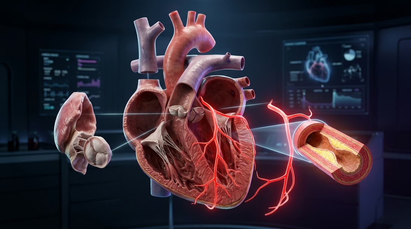
The human heart is an electromechanical marvel, contracting over 100,000 times per day to circulate roughly 7,500 liters of blood through a 100,000-kilometer vascular network. Yet, beneath its macroscopic muscular rhythm lies a fragile balance of microvascular perfusion and hydrodynamic wall stress. When coronary perfusion is compromised by sub-endothelial lipid accumulation, the consequences are immediate, progressive, and frequently catastrophic.
To truly master cardiovascular physiology, one cannot rely solely on static two-dimensional textbook diagrams. Modern clinical medicine requires understanding how macroscopic chamber kinematics interact directly with microscopic vascular pathology.
In this clinical masterclass, we deconstruct the four-chamber human heart, trace the anatomical branching of the coronary arterial tree, examine the fluid mechanics of atrioventricular and semilunar valves, and explore the cellular progression of atherosclerotic plaque rupture using our interactive 3D WebGL workstation.
Interactive 3D Studio: Explode Heart & Coronary Tree
Run exact formula simulations on NexProTools.
1. Macroscopic Chamber Architecture & Systolic Hemodynamics
The human heart is structured as two parallel, synchronized muscular pumps separated by the central cardiac septum. The right heart operates as a low-pressure volumetric circuit delivering deoxygenated blood to the pulmonary bed, while the left heart operates as a high-pressure resistive circuit ejecting oxygenated blood across systemic vascular resistance.
The Left Ventricular Powerhouse
The left ventricle (LV) is the primary mechanical engine of systemic circulation. In healthy adults, the left ventricular free wall measures between 8 mm and 12 mm in end-diastolic thickness�nearly three times thicker than the compliant right ventricular wall (3 mm to 5 mm).
| Physiological Metric | Left Ventricle (LV) | Right Ventricle (RV) | Clinical Significance | | :--- | :--- | :--- | :--- | | **Mean Systolic Pressure** | 110 - 130 mmHg | 20 - 25 mmHg | LV generates systemic perfusion pressure; RV preserves low pulmonary bed resistance | | **End-Diastolic Volume (EDV)** | 100 - 140 mL | 100 - 160 mL | Filling capacity index governing Frank-Starling preload stretching | | **End-Systolic Volume (ESV)** | 35 - 50 mL | 40 - 60 mL | Residual volume after ejection; elevates sharply in systolic pump failure | | **Resting Stroke Volume** | 65 - 85 mL | 65 - 85 mL | Strict volumetric equivalence between systemic and pulmonary circuits | | **Normal Ejection Fraction** | 55% - 70% | 45% - 60% | Primary clinical index of global contractile performance | | **Myocardial Wall Thickness** | 8 - 12 mm | 3 - 5 mm | LV wall is 3x thicker to normalize high Laplace systolic wall stress |
During isovolumetric contraction, the left ventricle generates rapid pressure rise (dP/dt > 1200 mmHg/s) to overcome aortic diastolic pressure (typically 80 mmHg). Once left ventricular cavity pressure exceeds aortic pressure, the aortic valve leaflets snap open, initiating rapid systolic ejection.
Laplace's Law and Myocardial Wall Stress
The mechanical stress experienced by individual cardiac myocytes is governed by the Law of Laplace for thick-walled spherical structures:
Where:
\sigmarepresents myocardial wall tension (force per unit cross-sectional area).Prepresents left ventricular intracavitary pressure (mmHg).rrepresents the internal radius of the ventricular cavity (cm).hrepresents ventricular myocardial wall thickness (cm).
This fundamental equation explains the divergence between physiological and pathological cardiac remodeling:
- Concentric Hypertrophy (Pressure Overload): In chronic systemic hypertension or aortic stenosis, elevated intracavitary pressure (
P) drives compensatory thickening of the muscular wall (h). By increasing wall thickness, the myocyte normalizes wall stress (\sigma), maintaining stroke volume at the expense of diastolic compliance. - Eccentric Hypertrophy & Dilatation (Volume Overload): In mitral regurgitation or dilated cardiomyopathy, end-diastolic radius (
r) progressively enlarges. Without a proportionate increase in wall thickness, systolic wall stress skyrockets, accelerating energetic exhaustion and clinical heart failure (HFrEF).
2. The Coronary Arterial Tree: Perfusion Territories & Dominance
Despite pumping millions of liters of blood through its interior chambers, the myocardium cannot extract oxygen or nutrients from the blood pooling inside its ventricles. The thick muscular walls require an external microvascular network: the Coronary Arteries.
The coronary arteries arise directly from the aortic root at the right and left sinuses of Valsalva, immediately superior to the aortic valve cusps.
``> CORONARY ARTERY TREE BRANCHING HIERARCHY
- Left Coronary System: Aortic Root (Left Sinus of Valsalva) $\to$ Left Main Coronary Artery (LMCA)
- $\to$ Left Anterior Descending (LAD): Septal perforators (anterior 2/3 septum) + Diagonal branches (anterior LV free wall & apex).
- $\to$ Left Circumflex (LCx): Obtuse Marginal (OM) branches (lateral and posterior LV wall + left atrium).
- Right Coronary System: Aortic Root (Right Sinus of Valsalva) $\to$ Right Coronary Artery (RCA)
- $\to$ Acute Marginal branches (RV free wall) + SA / AV Nodal arteries (cardiac conduction pacemakers).
- $\to$ Posterior Descending Artery (PDA) (inferior LV wall and posterior 1/3 septum in 85% right-dominant individuals).``
The Left Coronary System
The Left Main Coronary Artery (LMCA, typically 3.5 mm to 5.0 mm in caliber) courses between the pulmonary trunk and the left atrial appendage for 5 mm to 20 mm before bifurcating into two critical branches:
1. Left Anterior Descending Artery (LAD)
Coursing down the anterior interventricular sulcus toward the cardiac apex, the LAD is universally recognized as the most critical arterial vessel in human physiology�clinically referred to as the "Widowmaker":
- Perfusion Territory: Supplies the anterior two-thirds of the interventricular septum (via septal perforator branches), the anterior left ventricular free wall, the anterolateral papillary muscle, and the cardiac apex.
- Hemodynamic Impact: Proximal occlusion of the LAD deprives over 45% of total left ventricular myocardial mass of oxygen, precipitating acute cardiogenic shock, malignant ventricular arrhythmias (VT/VF), and death within minutes without emergency percutaneous coronary intervention (
PCI).
2. Left Circumflex Artery (LCx)
Branches at nearly a 90-degree angle from the LMCA to travel posterior-laterally within the coronary atrioventricular sulcus:
- Perfusion Territory: Gives off obtuse marginal (
OM) branches that supply the lateral and posterior left ventricular free walls, as well as the left atrium.
The Right Coronary System (RCA)
Arising from the anterior right aortic sinus, the RCA descends along the right atrioventricular groove toward the crux of the heart:
- Conduction System Perfusion: In 60% of humans, the sinus node artery branches from the proximal RCA; in 90% of humans, the atrioventricular (
AV) nodal artery branches from the distal RCA. Consequently, inferior myocardial infarctions caused by acute RCA thrombosis are frequently accompanied by severe sinus bradycardia or complete heart block. - Coronary Dominance: Coronary dominance is defined strictly by which artery gives rise to the Posterior Descending Artery (
PDA), which supplies the posterior third of the interventricular septum:- Right-Dominant (85% of population): The PDA branches from the distal RCA.
- Left-Dominant (8% to 10% of population): The PDA branches from the distal terminal circumflex (
LCx). - Co-Dominant (5% to 7% of population): Branches from both the RCA and LCx contribute to posterior septal perfusion.
| Vessel Name | Normal Lumen Caliber | Primary Myocardial Territory | Clinical Occlusion Outcome | | :--- | :--- | :--- | :--- | | **Left Main (LMCA)** | 4.0 - 5.5 mm | Global Left Ventricular Engine | Cardiogenic collapse; urgent CABG indication | | **Left Anterior Descending (LAD)** | 3.0 - 4.2 mm | Anterior Wall, 2/3 Septum, Apex | Widowmaker transmural anterior STEMI | | **Left Circumflex (LCx)** | 2.5 - 3.8 mm | Lateral Free Wall, Left Atrium | Lateral / Posterior STEMI (frequently silent on 12-lead ECG) | | **Right Coronary (RCA)** | 3.2 - 4.5 mm | RV Free Wall, SA/AV Nodes, Posterior Septum | Inferior STEMI with bradycardia / complete AV heart block | | **Posterior Descending (PDA)** | 1.8 - 2.8 mm | Inferior Wall, Posterior 1/3 Septum | Inferior wall ischemia; diaphragmatic chest pain |
3. Valvular Biomechanics & Turbulent Flow Dynamics
Cardiac valves are passive, non-muscular structures that ensure strict unidirectional blood transit across the cardiovascular loop. Their cyclic opening and closing are driven entirely by trans-valvular pressure gradients.
| Cardiac Phase | Atrioventricular Valves (Mitral & Tricuspid) | Semilunar Valves (Aortic & Pulmonic) | Intracavitary Hemodynamics | | :--- | :--- | :--- | :--- | | **Ventricular Systole** | **CLOSED** (Papillary muscles tense chordae tendineae to prevent atrial prolapse) | **OPEN** (Ventricular pressures exceed 80 mmHg aortic / 10 mmHg pulmonary diastolic pressures) | Rapid ejection of 70 mL stroke volume into aorta and pulmonary trunk | | **Ventricular Diastole** | **OPEN** (Passive early filling followed by atrial systole atrial kick) | **CLOSED** (Aortic and pulmonary root elastic recoil closes fibroelastic cusps) | Coronary artery perfusion occurs predominantly during this relaxation phase |
The Mitral (Bicuspid) Valve Complex
Guarding the left atrioventricular orifice, the mitral valve consists of five anatomically integrated components: the fibrous annulus, anterior and posterior leaflets, chordae tendineae, and papillary muscles:
- Anterior Leaflet: Large, mobile, semi-circular leaflet that forms the posterior boundary of the Left Ventricular Outflow Tract (
LVOT). - Posterior Leaflet: Scalloped into three distinct anatomical segments (
P1,P2,P3), anchored directly to the posterior annulus. - Subvalvular Apparatus: Primary, secondary, and tertiary chordae tendineae originate from the anterolateral and posteromedial papillary muscles, inserting into the ventricular aspect of the leaflets. During ventricular systole, papillary muscle contraction exerts downward tension on the chordae, counterbalancing ventricular pressure and preventing leaflet prolapse into the left atrium.
The Aortic Semilunar Valve
Composed of three symmetrical fibroelastic cusps (Right Coronary Cusp, Left Coronary Cusp, and Non-Coronary Cusp) seated within the aortic root:
- Sinuses of Valsalva: The anatomical bulges behind each aortic cusp generate fluid eddy currents (vortices) during peak systolic ejection. These hydrodynamic vortices prevent the thin valve leaflets from striking the aortic wall and occluding the coronary ostia.
- Aortic Stenosis Fluid Mechanics: In calcific aortic stenosis, progressive fibrocalcific remodeling restricts valve cusp mobility, reducing the normal valve orifice area from
3.0 - 4.0 cm�down to< 1.0 cm�(severe stenosis). According to the Gorlin formula, fluid jet velocity across the narrowed orifice exceeds4.0 m/s, producing extreme systolic pressure gradients (> 40 mmHgmean gradient) and turbulent shear stress.
4. The Pathobiology of Atherosclerosis: From ApoB Retention to Plaque Rupture
Atherosclerosis is neither an inevitable consequence of aging nor a simple passive accumulation of inert fat. It is a chronic, non-resolving inflammatory disease of the arterial wall driven by the sub-endothelial retention of apolipoprotein B (ApoB)-containing lipoproteins.
``> CHRONOLOGICAL PROGRESSION OF CORONARY ATHEROMA
- ApoB Particle Retention: Circulating atherogenic lipoproteins cross the endothelium and bind electrostatically to intimal proteoglycans.
- Oxidation & Monocyte Recruitment: Entrapped LDL oxidizes into ox-LDL, triggering VCAM-1 and MCP-1 release to recruit circulating monocytes.
- Foam Cell Accumulation: Scavenger receptors (CD36, SR-A1) engulf ox-LDL unregulated, transforming macrophages into lipid-engorged foam cells.
- Fibrous Cap Thinning & Rupture: Defective efferocytosis produces a necrotic core; macrophage MMPs digest collagen, precipitating fibrous cap fracture.``
Stage 1: Endothelial Permeability and Particle Entrapment
The healthy arterial wall consists of three concentric layers:
- Tunica Intima: Single layer of endothelial cells resting upon a basal lamina and extracellular proteoglycan matrix.
- Tunica Media: Concentric layers of vascular smooth muscle cells (
VSMCs) regulating vasomotor tone. - Tunica Adventitia: Fibroblast collagen envelope containing the microvascular vasa vasorum and autonomic nerve fibers.
Atherogenesis initiates when circulating atherogenic lipoproteins (ApoB-100, predominantly LDL, Lp(a), and remnant particles) cross the compromised endothelial barrier at sites of disturbed, non-laminar shear stress�such as coronary bifurcations.
Once inside the tunica intima, the positively charged lysine and arginine residues of ApoB-100 bind electrostatically to negatively charged glycosaminoglycans on sub-endothelial proteoglycans (biglycan and decorin). This mechanical entrapment sequesters the particles within the arterial wall, preventing their egress back into circulation.
Stage 2: Oxidative Modification and Macrophage Infiltration
Trapped lipoproteins undergo enzymatic and oxidative modifications, transforming into oxidized LDL (ox-LDL). Ox-LDL acts as a potent pro-inflammatory danger-associated molecular pattern (DAMP):
- Adhesion Molecule Induction: Endothelial cells upregulate Vascular Cell Adhesion Molecule-1 (
VCAM-1) and Intercellular Adhesion Molecule-1 (ICAM-1). - Monocyte Recruitment: Circulating monocytes tether to the activated endothelium, roll, and extravasate into the intima guided by Monocyte Chemoattractant Protein-1 (
MCP-1 / CCL2). - Scavenger Receptor Ingestion: Within the intima, monocytes differentiate into tissue macrophages. Unlike physiological LDL receptors, macrophage scavenger receptors (
SR-A1andCD36) do not downregulate in response to intracellular cholesterol excess. Macrophages uncontrollably ingest ox-LDL particles until they become lipid-engorged Foam Cells.
| Molecular Stage | Biochemical Drivers | Cellular Effect | Pathological Consequence | | :--- | :--- | :--- | :--- | | **1. Sub-Endothelial Transcytosis** | Elevated circulating ApoB particle concentration | Infiltration into tunica intima at arterial branch points | Particle accumulation in extracellular matrix | | **2. Proteoglycan Entrapment** | Biglycan and decorin negatively charged GAGs | Electrostatic binding to positive ApoB-100 lysine/arginine residues | Irreversible retention within vascular wall | | **3. Oxidative Modification** | Reactive oxygen species (ROS), myeloperoxidase | Transition into oxidized LDL (ox-LDL) | Triggers pro-inflammatory DAMP immune signaling | | **4. Scavenger Ingestion** | Macrophage CD36 and SR-A1 receptors | Unregulated phagocytosis without negative feedback | Macrophage transforms into Foam Cell | | **5. Secondary Necrosis** | Apoptotic failure (defective efferocytosis) | Spilling of free cholesterol crystals and toxic oxysterols | Formation of destabilizing Lipid-Rich Necrotic Core |
Stage 3: Necrotic Core Formation and Fibrous Cap Attenuation
As foam cells become overwhelmed by cholesterol, they undergo programmed cell death (apoptosis). In early lesions, specialized efferocytosis mechanisms clear apoptotic remnants. However, as the lesion matures:
- Defective Efferocytosis: Dying macrophages undergo secondary necrosis, spilling free cholesterol, toxic oxysterols, and intracellular debris into the extracellular space.
- The Necrotic Core: This accumulated lipid debris forms a cellular-free, destabilizing Lipid-Rich Necrotic Core (
LRNC). - Fibrous Cap Synthesis: Neighboring vascular smooth muscle cells (
VSMCs) migrate from the tunica media into the intima under platelet-derived growth factor (PDGF) signaling. These VSMCs synthesize extracellular matrix, creating a structural collagen-rich Fibrous Cap that shields the thrombogenic necrotic core from the flowing bloodstream.
5. Vulnerable Plaque Mechanics & Acute Coronary Thrombosis
Not all atherosclerotic plaques are created equal. Ironically, high-grade, calcified plaques that cause 80% - 90% stable luminal narrowing frequently do not cause sudden fatal heart attacks. Instead, over 70% of acute myocardial infarctions arise from non-obstructive (< 50% stenosis) but dynamically unstable lesions termed Thin-Cap Fibroatheromas (TCFA).
| Morphological Feature | Vulnerable Thin-Cap Fibroatheroma (TCFA) | Stable Fibrous Plaque | | :--- | :--- | :--- | | **Fibrous Cap Thickness** | **< 65 micrometers** (Mechanically fragile) | **> 150 micrometers** (Dense, thick collagen shield) | | **Necrotic Core Volume** | Large (> 25% of total plaque volume) | Small (< 10% of total lesion volume) | | **Macrophage Inflammation** | Intense / Activated at plaque shoulders (High MMPs) | Minimal / Quiescent cellular infiltration | | **VSMC Smooth Muscle Content**| Severely depleted (Reduced collagen repair) | Abundant (Active ongoing extracellular matrix synthesis) | | **Microcalcification Pattern** | Spotty / Speckled microcalcifications (< 50 �m) | Dense coalescent sheet calcification (Stable CAC) | | **Intraplaque Neovascularization**| Dense, leaky immature vasa vasorum with micro-hemorrhage| Absent or matured microvasculature | | **Primary Clinical Event** | **Acute Transmural STEMI / Sudden Cardiac Death** | **Stable exertional angina (Predictable threshold)** |
The Biomechanics of Fibrous Cap Rupture
A fibrous cap ruptures when hemodynamic tensile peak wall stress exceeds the tensile material strength of the cap tissue:
Where:
t_{\text{cap}}is fibrous cap thickness. Whent_{\text{cap}} < 65 �m, the cap lacks sufficient mechanical tensile resistance.- Macrophages at the vulnerable shoulder regions of the plaque secrete Matrix Metalloproteinases (
MMP-1,MMP-8,MMP-13collagenases) andMMP-2,MMP-9gelatinases. These enzymes cleave triple-helical Type I and Type III collagen strands, actively digesting the cap from within. - Interstitial microcalcifications (spotty calcium deposits
< 50 �m) act as local mechanical stress concentrators, amplifying tensile forces along the cap interface by up to 500%.
``> ACUTE THROMBOGENESIS CASCADE UPON CAP FRACTURE
- Fibrous Cap Fissure: Tensile hemodynamic peak wall stress exceeds cap material strength (t_cap < 65 �m$).
- Tissue Factor Exposure: Flowing blood directly contacts necrotic core tissue factor (TF) and sub-endothelial collagen.
- Platelet Adhesion: Platelets anchor to von Willebrand factor via glycoprotein Ib-IX-V and collagen via GP VI receptors.
- Platelet Activation & Degranulation: Release of thromboxane A2 ($) and ADP recruits circulating platelets.
- Fibrin Mesh Cross-Linking: Conformational activation of GP IIb/IIIa receptors binds fibrinogen, assembling an occlusive intraluminal red thrombus.``
When the cap ruptures, blood inside the arterial lumen directly contacts the highly pro-thrombotic lipid core. Tissue factor (TF) binds to Factor VIIa, triggering the extrinsic coagulation cascade, while sub-endothelial von Willebrand factor (vWF) anchors circulating platelets via glycoprotein Ib-IX-V. Within seconds, platelets aggregate via activated GP IIb/IIIa cross-linked by fibrinogen, producing an occlusive intraluminal thrombus that abruptly halts all downstream blood flow.
6. Clinical Risk Stratification: Laboratory Biomarkers & Imaging
Evaluating cardiovascular risk requires moving beyond crude total cholesterol figures toward precision circulating biomarkers and direct non-invasive anatomical imaging.
| Biomarker | Optimal Target | Clinical Predictive Value | Underlying Pathological Mechanism | | :--- | :--- | :--- | :--- | | **Apolipoprotein B (ApoB)** | **< 60 mg/dL** | Absolute quantification of atherogenic particle count | Measures all circulating atherogenic particles (LDL, VLDL, IDL, Lp(a)) | | **Lipoprotein(a) [Lp(a)]** | **< 30 mg/dL** | Highly atherogenic, pro-thrombotic genetic risk driver | Competes with plasminogen, accelerating thrombosis and intimal oxidation | | **High-Sensitivity CRP (hs-CRP)**| **< 0.8 mg/L** | Vascular endothelial and systemic arterial inflammation | Downstream marker of IL-6 and macrophage activation within plaque shoulders | | **High-Sensitivity Troponin I** | **< 5 ng/L** | Detection of subclinical myocyte ischemic micro-necrosis | Sarcomeric protein released during transient coronary perfusion mismatches | | **Coronary Calcium Score (CAC)** | **0 Agatston Units**| Direct quantification of lifetime calcified plaque burden | Computed tomography scoring of fibrocalcific arterial remodeling |
Apolipoprotein B (ApoB) vs LDL-Cholesterol
Standard lipid panels calculate LDL-C (the mass of cholesterol carried within LDL particles). However, cholesterol mass per particle varies dramatically:
- In individuals with insulin resistance, obesity, or metabolic syndrome, LDL particles are depleted of cholesterol esters and enriched with triglycerides, creating Small Dense LDL (
sdLDL). - While their calculated
LDL-Cmay appear deceptively normal, their actual particle concentration is dangerously elevated. - Because every atherogenic particle (VLDL, IDL, LDL, Lp(a)) contains exactly one molecule of
ApoB-100, measuring circulatingApoBquantifies the absolute particle concentration invading the arterial wall.
Explore our dedicated ApoB Cardiovascular Risk Calculator to evaluate particle retention kinetics.
High-Sensitivity Troponin I (hs-cTnI)
Cardiac troponin I is a regulatory structural protein of the contractile sarcomere. With fifth-generation high-sensitivity assays, clinicians can detect picogram-per-milliliter concentrations of troponin released during transient micro-ischemia, subclinical plaque fissure, or myocyte apoptosis�identifying elevated cardiovascular mortality risk years before an overt clinical heart attack occurs.
7. Interactive 3D Disassembly & Anatomical Inspection Protocol
To maximize your learning when exploring our 3D Human Anatomy Studio, follow this structured four-step clinical exploration protocol:
Step 1: Anterior Wall Disassembly (Exploded View: 0% to 50%)
- Rotate the heart model in 3D space to inspect the natural orientation of the anterior interventricular sulcus.
- Drag the Layer Disassembly Slider to
35%. - Observe how the anterior myocardial shell lifts away, revealing the interior cavity of the left ventricle, the muscular interventricular septum, and the delicate leaflets of the mitral and tricuspid valves.
Step 2: Great Vessel Outflow Orientation
- Elevate the camera angle to inspect the superior aspect of the cardiac base.
- Trace the path of the Ascending Aorta as it arches superiorly, giving off the brachiocephalic trunk, left common carotid, and left subclavian arteries.
- Observe the anterior crossing of the pulmonary trunk as it bifurcates beneath the aortic arch.
Step 3: Coronary Perfusion Tracing
- Rotate the heart to trace the Left Anterior Descending (
LAD) artery from its origin behind the pulmonary trunk down to the apex. - Locate the Right Coronary Artery (
RCA) running in the right atrioventricular groove. - Click on the 3D annotation pins to view the associated Latin terminology, ICD-10 diagnostic classifications, and clinical laboratory biomarkers.
Step 4: Pathology Mode Activation (Atherosclerosis Simulation)
- Toggle Clinical Pathology Simulation to
Active. - Observe the procedural cross-sectional cutaway of the proximal LAD.
- Increase the Severity Slider from
20%to90%:- Watch the golden-yellow lipid pool expand within the sub-endothelial space.
- Note the appearance of white, glistening calcified crystals within the necrotic core.
- Observe the progressive narrowing (stenosis) of the functional red arterial lumen, demonstrating the hemodynamic transition from normal laminar flow to ischemic critical stenosis.
8. Summary & Key Takeaways
CLINICAL TAKEAWAYS: 3D CARDIOVASCULAR ANATOMY
- The Left Ventricle operates at three times the muscular wall thickness of the right ventricle, with myocardial wall tension strictly governed by Laplace's Law (
\sigma = (P \cdot r) / (2h)).- The LAD Artery ("Widowmaker") supplies over 45% of left ventricular myocardium; proximal occlusion leads to massive transmural anterior STEMI and rapid cardiogenic collapse.
- Atherosclerosis Initiates not with passive fat accumulation, but with the sub-endothelial mechanical entrapment of
ApoB-containing lipoproteins binding to intimal proteoglycans.- Plaque Rupture is primarily caused by matrix metalloproteinases cleaving collagen within thin fibrous caps (
< 65 �m), exposing thrombogenic tissue factor to the bloodstream.- Precision Risk Assessment mandates measuring circulating
ApoBparticle count alongside High-Sensitivity Troponin (hs-cTnI) and Coronary Artery Calcium (CAC) scoring.
Mastering human anatomy requires spatial intuition. We invite you to explore the Interactive 3D Human Anatomy Workstation to inspect these anatomical structures in real-time WebGL 3D.